Mallio Carlo Augusto, Tomarchio Valeria, Pulcini Francesco, Verducci Edoardo, Bernetti Caterina, Tafuri Maria Antonietta, Greco Federico, Rigacci Luigi, Zobel Bruno Beomonte, Annibali Ombretta
Fondazione Policlinico Universitario Campus Bio-Medico, Via Alvaro del Portillo, 200, 00128 Roma, Italy.
Research Unit Diagnostic Imaging, Fondazione Policlinico Universitario Campus Bio-Medico, Via Alvaro del Portillo, 200, 00128 Roma, Italy.
Hematol Rep. 2024 Oct 17;16(4):624-635. doi: 10.3390/hematolrep16040061.
The aim of this study was to evaluate the impact of trabecular attenuation of the L1 vertebral body in low-dose CT in adult patients with multiple myeloma (MM), smoldering multiple myeloma (SMM), and monoclonal gammopathy of undetermined significance (MGUS).
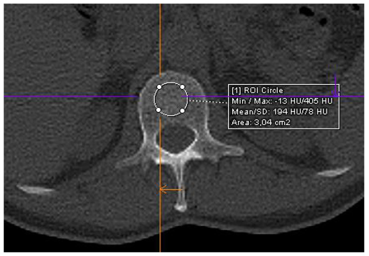
The study population consisted of 22 patients with MGUS and 51 consecutive patients with newly diagnosed MM (SMM, = 21; symptomatic MM, = 36). CT scans were conducted using a 128-slice CT scanner (Somatom go.Top, Siemens, Munich, Germany). Low-dose whole-body CT scans were performed at a single time point for each patient. Trabecular bone density values were obtained by defining regions of interest on non-contrast images at the level of L1 vertebra. A threshold of = 0.05 was applied to determine statistical significance.
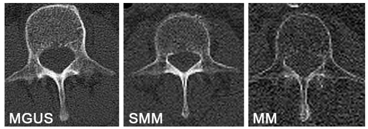
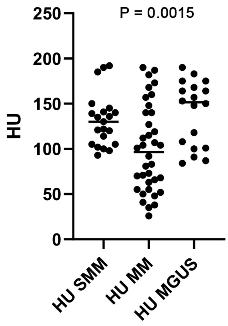
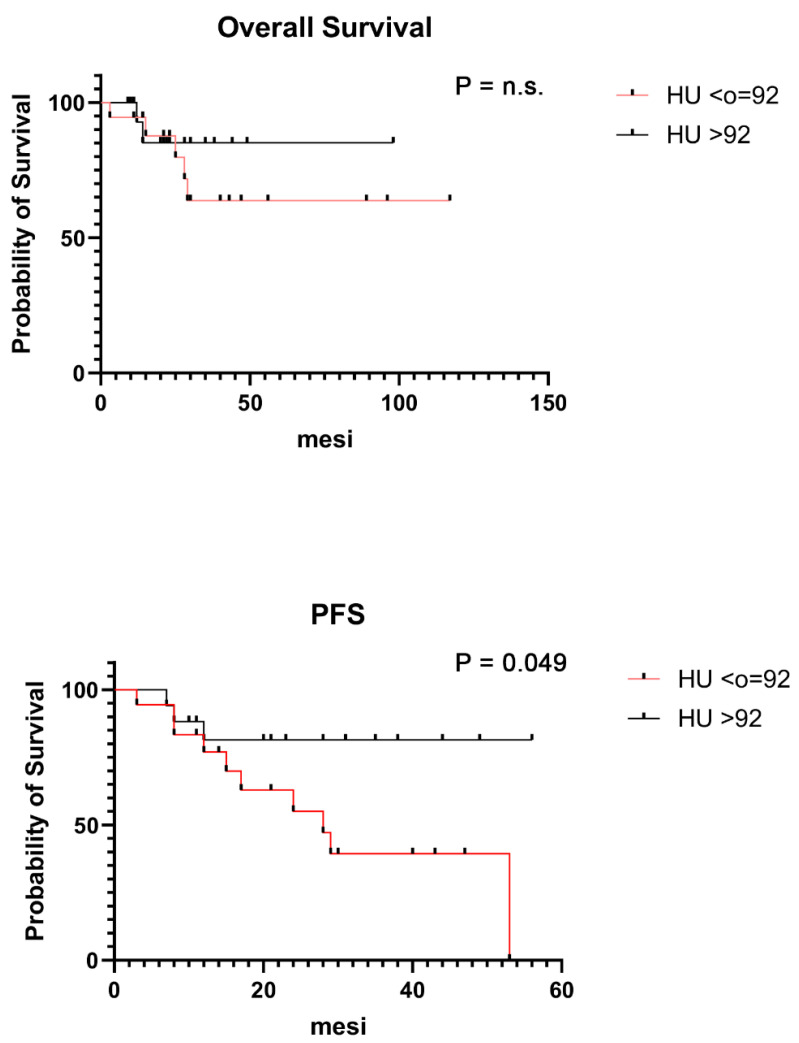
The median Hounsfield unit (HU) value in patients with MGUS, SMM, and MM was 148 HU (range 81-190), 130 HU (range 93-193), and 92 HU (range 26-190), respectively, with a statistically significant difference between the groups ( = 0.0015). Patients with HU values ≤ 92 had lower progression-free survival with statistically significant differences compared to the group with HU values > 92 ( < 0.0499).
This is the earliest evidence of the importance of evaluating L1 attenuation values in low-dose CT images in patients with MGUS, SMM, and MM. Further prospective studies could contribute to reinforcing these results and exploring the clinical applicability and generalization of L1 attenuation values in low-dose whole-body CT scans in routine clinical practice.
本研究旨在评估低剂量CT中L1椎体小梁衰减对成年多发性骨髓瘤(MM)、冒烟型多发性骨髓瘤(SMM)和意义未明的单克隆丙种球蛋白病(MGUS)患者的影响。
研究人群包括22例MGUS患者和51例新诊断的MM患者(SMM,21例;有症状MM,36例)。使用128层CT扫描仪(Somatom go.Top,西门子,慕尼黑,德国)进行CT扫描。对每位患者在单个时间点进行低剂量全身CT扫描。通过在L1椎体水平的非增强图像上定义感兴趣区域来获得小梁骨密度值。采用α = 0.05的阈值来确定统计学显著性。
MGUS、SMM和MM患者的中位亨氏单位(HU)值分别为148 HU(范围81 - 190)、130 HU(范围93 - 193)和92 HU(范围26 - 190),组间差异有统计学意义(P = 0.0015)。HU值≤92的患者无进展生存期较低,与HU值>92的组相比差异有统计学意义(P < 0.0499)。
这是关于评估MGUS、SMM和MM患者低剂量CT图像中L1衰减值重要性的最早证据。进一步的前瞻性研究可能有助于强化这些结果,并探索L1衰减值在常规临床实践中低剂量全身CT扫描的临床适用性和普遍性。