• 文献检索
  • 文档翻译
  • 深度研究
  • 学术资讯
  • Suppr Zotero 插件Zotero 插件
  • 邀请有礼
  • 套餐&价格
  • 历史记录
应用&插件
Suppr Zotero 插件Zotero 插件浏览器插件Mac 客户端Windows 客户端微信小程序
定价
高级版会员购买积分包购买API积分包
服务
文献检索文档翻译深度研究API 文档MCP 服务
关于我们
关于 Suppr公司介绍联系我们用户协议隐私条款
关注我们

Suppr 超能文献

核心技术专利:CN118964589B侵权必究
粤ICP备2023148730 号-1Suppr @ 2026

文献检索

告别复杂PubMed语法,用中文像聊天一样搜索,搜遍4000万医学文献。AI智能推荐,让科研检索更轻松。

立即免费搜索

文件翻译

保留排版,准确专业,支持PDF/Word/PPT等文件格式,支持 12+语言互译。

免费翻译文档

深度研究

AI帮你快速写综述,25分钟生成高质量综述,智能提取关键信息,辅助科研写作。

立即免费体验

从锥鞭毛体到锥鞭毛体:通过超微结构扩展显微镜分析[具体内容]的单向细胞内旅程 。 (注:原文中“Analyzing the One-Way Intracellular Journey of by Ultrastructure Expansion Microscopy.”里“of”后面缺少具体所指内容)

From Trypomastigotes to Trypomastigotes: Analyzing the One-Way Intracellular Journey of by Ultrastructure Expansion Microscopy.

作者信息

Tomasina Ramiro, González Fabiana C, Cabrera Andrés, Basmadjián Yester, Robello Carlos

机构信息

Laboratorio de Interacciones Hospedero Patógeno, Institut Pasteur de Montevideo, Montevideo 11400, Uruguay.

Unidad Académica de Parasitología y Micología, Facultad de Medicina, Universidad de la República, Montevideo 11550, Uruguay.

出版信息

Pathogens. 2024 Oct 2;13(10):866. doi: 10.3390/pathogens13100866.

DOI:10.3390/pathogens13100866
PMID:39452737
原文链接:https://pmc.ncbi.nlm.nih.gov/articles/PMC11510640/
Abstract

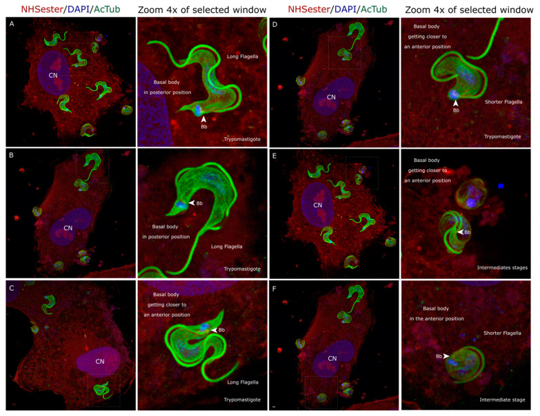
The protozoan parasite is the causative agent of Chagas disease, also called American trypanosomiasis. This neglected tropical disease affects millions of individuals across the Americas. To complete its life cycle, parasitizes both vertebrate hosts and its vector, commonly known as the 'kissing bug'. The parasite's survival and proliferation strategies are driven by the diverse environments it encounters. Despite being described by Carlos Chagas in 1909, significant knowledge gaps persist regarding the parasite's various life forms and adaptive capabilities in response to environmental cues. In this study, we employed Ultrastructure Expansion Microscopy to explore the intricate journey of within the host cell. Upon entry into the host cell, trypomastigotes undergo folding, resulting in intermediate forms characterized by a rounded cell body, anterior positioning of basal bodies, and a shortened flagellum. The repositioning of basal bodies and the kinetoplast and the shortening of the flagella mark the culmination of intracellular amastigogenesis. Furthermore, we analyzed intracellular trypomastigogenesis, identifying discrete intermediate forms, including leaf-shaped stages and epimastigote-like forms, which suggests a complex differentiation process. Notably, we did not observe any dividing intracellular epimastigotes, indicating that these may be non-replicative forms within the host cell. Our detailed examination of amastigote cell division revealed semi-closed nuclear mitosis, with mitotic spindle formation independent of basal bodies. This study provides new insights into the morphological and cytoskeletal changes during the intracellular stages of , providing a model for understanding the dynamics of intracellular amastigogenesis and trypomastigogenesis.

摘要

这种原生动物寄生虫是恰加斯病(也称为美洲锥虫病)的病原体。这种被忽视的热带疾病影响着美洲数百万人口。为了完成其生命周期,该寄生虫寄生于脊椎动物宿主及其传播媒介(通常称为“接吻虫”)。寄生虫的生存和增殖策略受其所处不同环境的驱动。尽管卡洛斯·恰加斯在1909年就对其进行了描述,但关于该寄生虫的各种生命形式以及对环境线索的适应能力仍存在重大知识空白。在本研究中,我们采用超微结构扩展显微镜来探索该寄生虫在宿主细胞内的复杂历程。进入宿主细胞后,锥鞭毛体发生折叠,形成中间形态,其特征为细胞体呈圆形、基体位于前部且鞭毛缩短。基体和动基体的重新定位以及鞭毛的缩短标志着细胞内无鞭毛体生成的完成。此外,我们分析了细胞内锥鞭毛体生成过程,识别出离散的中间形态,包括叶状阶段和类上鞭毛体形态,这表明存在一个复杂的分化过程。值得注意的是,我们未观察到任何正在分裂的细胞内上鞭毛体,这表明这些可能是宿主细胞内的非复制形式。我们对无鞭毛体细胞分裂的详细检查揭示了半封闭的核有丝分裂,有丝分裂纺锤体的形成独立于基体。本研究为该寄生虫细胞内阶段的形态和细胞骨架变化提供了新见解,为理解细胞内无鞭毛体生成和锥鞭毛体生成的动态过程提供了一个模型。

https://cdn.ncbi.nlm.nih.gov/pmc/blobs/2c17/11510640/27c8efbffdf9/pathogens-13-00866-g007.jpg
https://cdn.ncbi.nlm.nih.gov/pmc/blobs/2c17/11510640/c6d214cb2718/pathogens-13-00866-g001.jpg
https://cdn.ncbi.nlm.nih.gov/pmc/blobs/2c17/11510640/e469b75749ed/pathogens-13-00866-g002.jpg
https://cdn.ncbi.nlm.nih.gov/pmc/blobs/2c17/11510640/c12e7ce1d3c9/pathogens-13-00866-g003.jpg
https://cdn.ncbi.nlm.nih.gov/pmc/blobs/2c17/11510640/c936706b34e3/pathogens-13-00866-g004.jpg
https://cdn.ncbi.nlm.nih.gov/pmc/blobs/2c17/11510640/b624a7b0bb7b/pathogens-13-00866-g005.jpg
https://cdn.ncbi.nlm.nih.gov/pmc/blobs/2c17/11510640/4c2e28e7c1b0/pathogens-13-00866-g006.jpg
https://cdn.ncbi.nlm.nih.gov/pmc/blobs/2c17/11510640/27c8efbffdf9/pathogens-13-00866-g007.jpg

相似文献

1
From Trypomastigotes to Trypomastigotes: Analyzing the One-Way Intracellular Journey of by Ultrastructure Expansion Microscopy.从锥鞭毛体到锥鞭毛体:通过超微结构扩展显微镜分析[具体内容]的单向细胞内旅程 。 (注:原文中“Analyzing the One-Way Intracellular Journey of by Ultrastructure Expansion Microscopy.”里“of”后面缺少具体所指内容)
Pathogens. 2024 Oct 2;13(10):866. doi: 10.3390/pathogens13100866.
2
Parasite-Mediated Remodeling of the Host Microfilament Cytoskeleton Enables Rapid Egress of Trypanosoma cruzi following Membrane Rupture.寄生虫介导的宿主微丝细胞骨架重排使克氏锥虫能够在膜破裂后迅速逸出。
mBio. 2021 Jun 29;12(3):e0098821. doi: 10.1128/mBio.00988-21. Epub 2021 Jun 22.
3
Comprehensive glycoprofiling of the epimastigote and trypomastigote stages of Trypanosoma cruzi.全面糖基谱分析克氏锥虫的前鞭毛体和无鞭毛体阶段。
J Proteomics. 2017 Jan 16;151:182-192. doi: 10.1016/j.jprot.2016.05.034. Epub 2016 Jun 16.
4
The marsupial trypanosome Trypanosoma copemani is not an obligate intracellular parasite, although it adversely affects cell health.有袋动物锥体虫 Trypanosoma copemani 并非必需的细胞内寄生虫,尽管它会对细胞健康造成不利影响。
Parasit Vectors. 2018 Sep 20;11(1):521. doi: 10.1186/s13071-018-3092-1.
5
The thermal proteome stability profile of Trypanosoma cruzi in epimastigote and trypomastigote life stages.锥虫热蛋白组稳定性分析在其上皮样鞭毛体和动基体阶段。
J Proteomics. 2021 Sep 30;248:104339. doi: 10.1016/j.jprot.2021.104339. Epub 2021 Aug 2.
6
Revisiting the Trypanosoma cruzi metacyclogenesis: morphological and ultrastructural analyses during cell differentiation.重新审视克氏锥虫的循环形态发生:细胞分化过程中的形态和超微结构分析。
Parasit Vectors. 2018 Feb 6;11(1):83. doi: 10.1186/s13071-018-2664-4.
7
Morphological comparison of axenic amastigogenesis of trypomastigotes and metacyclic forms of Trypanosoma cruzi.克氏锥虫无鞭毛体生成及循环后期形态的体外形态学比较
Mem Inst Oswaldo Cruz. 2003 Jan;98(1):83-91. doi: 10.1590/s0074-02762003000100012. Epub 2003 Apr 9.
8
Trypanosoma cruzi: characterization of an intracellular epimastigote-like form.克氏锥虫:一种细胞内类上鞭毛体形式的特征描述。
Exp Parasitol. 1999 Aug;92(4):263-74. doi: 10.1006/expr.1999.4423.
9
Flagellar motility of Trypanosoma cruzi epimastigotes.克氏锥虫前鞭毛体的鞭毛运动
J Biomed Biotechnol. 2012;2012:520380. doi: 10.1155/2012/520380. Epub 2012 Jan 11.
10
Host cell actin remodeling in response to Trypanosoma cruzi: trypomastigote versus amastigote entry.宿主细胞肌动蛋白重塑对克氏锥虫的反应:锥鞭毛体与无鞭毛体的入侵。
Subcell Biochem. 2008;47:101-9. doi: 10.1007/978-0-387-78267-6_8.

引用本文的文献

1
Differential susceptibility of human placental trophoblasts to Trypanosoma cruzi infection.人胎盘滋养层细胞对克氏锥虫感染的易感性差异
Parasitol Res. 2025 Sep 16;124(9):104. doi: 10.1007/s00436-025-08552-7.
2
Mitochondrial DNA Structure in .线粒体DNA结构于……中 (原文不完整,翻译可能不太准确,需结合完整原文进一步完善)
Pathogens. 2025 Jan 14;14(1):73. doi: 10.3390/pathogens14010073.

本文引用的文献

1
Insights into the Cell Division of .对……细胞分裂的见解 。 你提供的原文似乎不完整,请补充完整以便我能准确翻译。
Microorganisms. 2023 Dec 28;12(1):61. doi: 10.3390/microorganisms12010061.
2
Atlas of intraerythrocytic development using expansion microscopy.使用扩展显微镜的红细胞内发育图谱。
Elife. 2023 Dec 18;12:RP88088. doi: 10.7554/eLife.88088.
3
Chagas disease.恰加斯病
Lancet. 2024 Jan 13;403(10422):203-218. doi: 10.1016/S0140-6736(23)01787-7. Epub 2023 Dec 7.
4
The hows and whys of amastigote flagellum motility in .无鞭毛阿米巴运动的方式和原因。
mBio. 2023 Aug 31;14(4):e0053123. doi: 10.1128/mbio.00531-23. Epub 2023 Jun 6.
5
Ultrastructural Expansion Microscopy in Three In Vitro Life Cycle Stages of Trypanosoma cruzi.锥虫克鲁兹氏体内三个体外生命周期阶段的超微结构扩张显微镜观察。
J Vis Exp. 2023 May 12(195). doi: 10.3791/65381.
6
3D FIB-SEM structural insights into the architecture of sub-pellicular microtubules of Trypanosoma cruzi epimastigotes.三维 FIB-SEM 结构研究克氏锥虫滋养体表膜下微管的结构。
Biol Cell. 2022 Aug;114(8):203-210. doi: 10.1111/boc.202100038. Epub 2022 May 3.
7
Revisiting the Role of Toxoplasma gondii ERK7 in the Maintenance and Stability of the Apical Complex.重新探讨刚地弓形虫 ERK7 在顶复体结构维持和稳定中的作用。
mBio. 2021 Oct 26;12(5):e0205721. doi: 10.1128/mBio.02057-21. Epub 2021 Oct 5.
8
The cytostome-cytopharynx complex of intracellular and extracellular amastigotes of Trypanosoma cruzi exhibit structural and functional differences.克氏锥虫细胞内和细胞外无鞭毛体的胞口-胞咽复合体表现出结构和功能上的差异。
Cell Microbiol. 2021 Sep;23(9):e13346. doi: 10.1111/cmi.13346. Epub 2021 May 18.
9
Expansion microscopy provides new insights into the cytoskeleton of malaria parasites including the conservation of a conoid.扩展显微镜为了解疟疾寄生虫的细胞骨架提供了新的视角,包括锥状体的保守性。
PLoS Biol. 2021 Mar 11;19(3):e3001020. doi: 10.1371/journal.pbio.3001020. eCollection 2021 Mar.
10
Ultrastructure expansion microscopy (U-ExM).超微结构扩展显微镜(U-ExM)。
Methods Cell Biol. 2021;161:57-81. doi: 10.1016/bs.mcb.2020.05.006. Epub 2020 Jul 1.