Centre for Microscopy, Characterisation and Analysis, The University of Western Australia, Crawley, Western Australia, 6009, Australia.
Central Analytical Research Facility, Queensland University of Technology, Brisbane, Queensland, 4000, Australia.
Parasit Vectors. 2018 Sep 20;11(1):521. doi: 10.1186/s13071-018-3092-1.
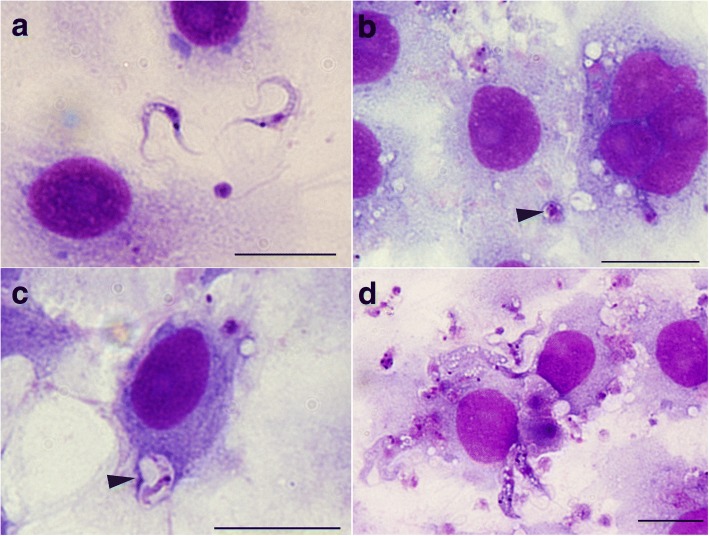
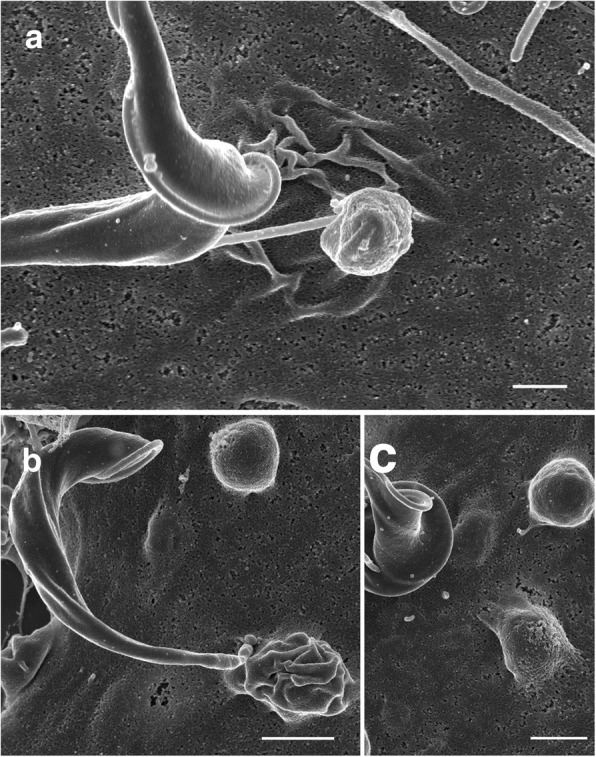
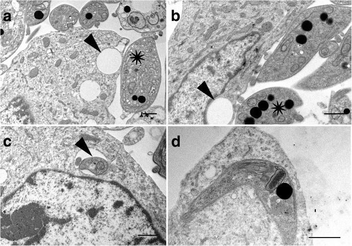
Trypanosoma cruzi invades and replicates inside mammalian cells, which can lead to chronic Chagas disease in humans. Trypanosoma copemani infects Australian marsupials and recent investigations indicate it may be able to invade mammalian cells in vitro, similar to T. cruzi. Here, T. cruzi 10R26 strain (TcIIa) and two strains of T. copemani [genotype 1 (G1) and genotype 2 (G2)] were incubated with marsupial cells in vitro. Live-cell time-lapse and fluorescent microscopy, combined with high-resolution microscopy (transmission and scanning electron microscopy) were used to investigate surface interactions between parasites and mammalian cells.
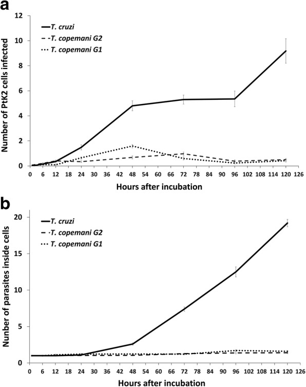
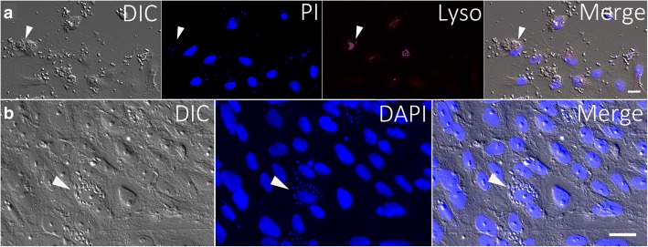
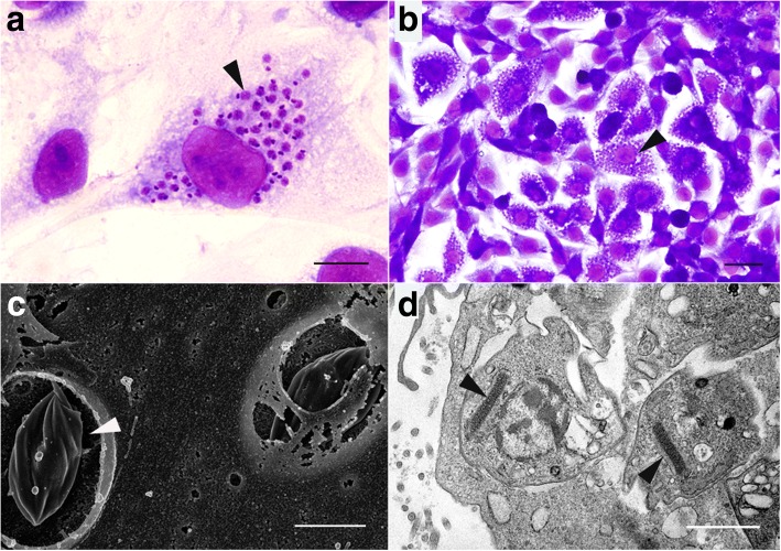
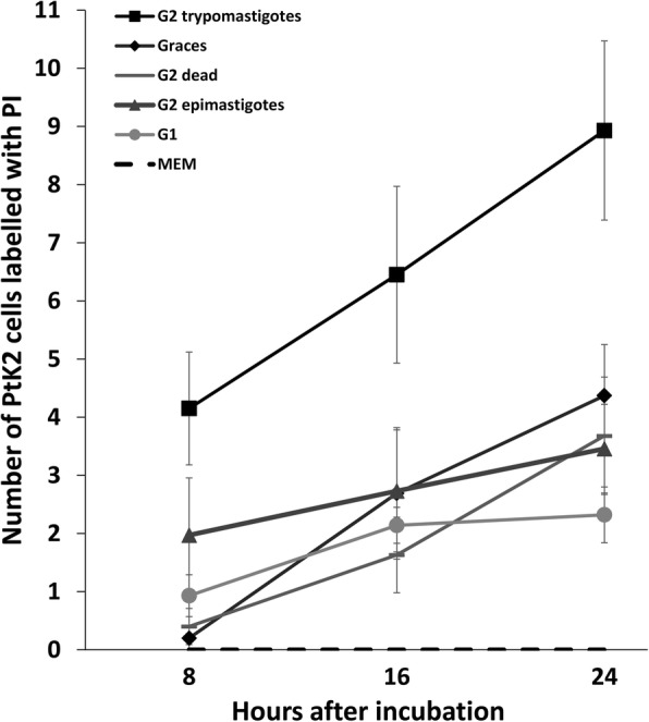
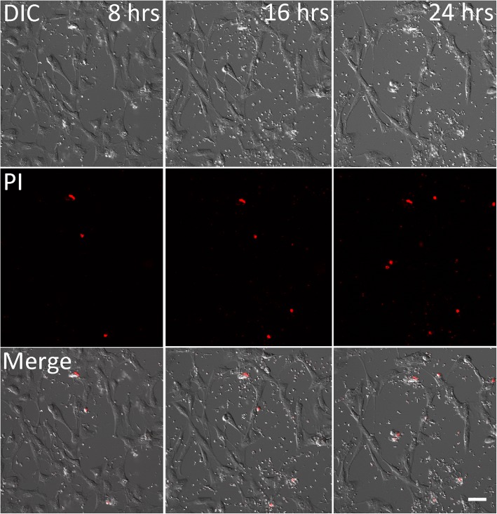
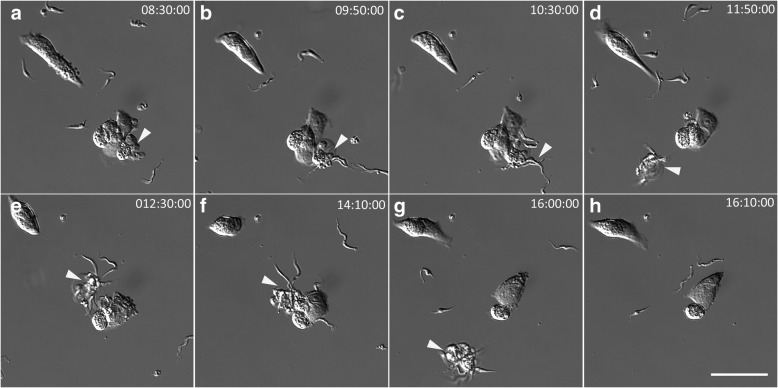
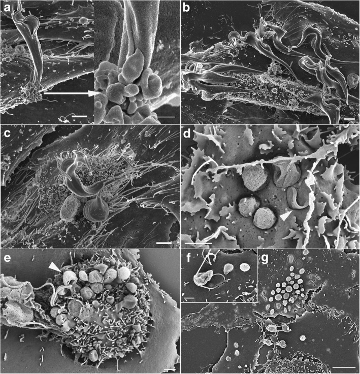
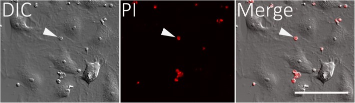
The number of parasites invading cells was significantly higher in T. cruzi compared to either genotype of T. copemani, between which there was no significant difference. While capable of cellular invasion, T. copemani did not multiply in host cells in vitro as there was no increase in intracellular amastigotes over time and no release of new trypomastigotes from host cells, as observed in T. cruzi. Exposure of host cells to G2 trypomastigotes resulted in increased host cell membrane permeability within 24 h of infection, and host cell death/blebbing was also observed. G2 parasites also became embedded in the host cell membrane.
Trypanosoma copemani is unlikely to have an obligate intracellular life-cycle like T. cruzi. However, T. copemani adversely affects cell health in vitro and should be investigated in vivo in infected host tissues to better understand this host-parasite relationship. Future research should focus on increasing understanding of the T. copemani life history and the genetic, physiological and ecological differences between different genotypes.
克氏锥虫入侵并在哺乳动物细胞内复制,可导致人类罹患慢性恰加斯病。克氏锥虫感染澳大利亚有袋类动物,最近的研究表明,它可能能够在体外入侵哺乳动物细胞,类似于克氏锥虫。在此,体外孵育了克氏锥虫 10R26 株(TcIIa)和两种克氏锥虫copemani 株[基因型 1(G1)和基因型 2(G2)]与有袋类细胞。使用活细胞延时和荧光显微镜,结合高分辨率显微镜(透射和扫描电子显微镜),研究了寄生虫与哺乳动物细胞之间的表面相互作用。
与任何一种基因型的克氏锥虫copemani 相比,克氏锥虫感染细胞的寄生虫数量明显更高,而两者之间没有显著差异。虽然能够进行细胞入侵,但克氏锥虫copemani 在体外宿主细胞中不会增殖,因为随着时间的推移,细胞内无鞭毛体没有增加,也没有新的锥虫从宿主细胞中释放,这与克氏锥虫的情况不同。宿主细胞暴露于 G2 锥虫后,在感染后 24 小时内宿主细胞膜通透性增加,并且观察到宿主细胞死亡/起泡。G2 寄生虫也嵌入宿主细胞膜中。
克氏锥虫copemani 不太可能像克氏锥虫那样具有强制性的细胞内生命周期。然而,克氏锥虫copemani 在体外对细胞健康有害,应该在感染宿主组织的体内进行研究,以更好地理解这种宿主-寄生虫关系。未来的研究应侧重于增加对克氏锥虫copemani 生活史以及不同基因型之间遗传、生理和生态差异的理解。