• 文献检索
  • 文档翻译
  • 深度研究
  • 学术资讯
  • Suppr Zotero 插件Zotero 插件
  • 邀请有礼
  • 套餐&价格
  • 历史记录
应用&插件
Suppr Zotero 插件Zotero 插件浏览器插件Mac 客户端Windows 客户端微信小程序
定价
高级版会员购买积分包购买API积分包
服务
文献检索文档翻译深度研究API 文档MCP 服务
关于我们
关于 Suppr公司介绍联系我们用户协议隐私条款
关注我们

Suppr 超能文献

核心技术专利:CN118964589B侵权必究
粤ICP备2023148730 号-1Suppr @ 2026

文献检索

告别复杂PubMed语法,用中文像聊天一样搜索,搜遍4000万医学文献。AI智能推荐,让科研检索更轻松。

立即免费搜索

文件翻译

保留排版,准确专业,支持PDF/Word/PPT等文件格式,支持 12+语言互译。

免费翻译文档

深度研究

AI帮你快速写综述,25分钟生成高质量综述,智能提取关键信息,辅助科研写作。

立即免费体验

增强双能 CT 胰腺实质期和延迟期扫描对胰腺癌的诊断价值。

Diagnostic Value of Contrast-Enhanced Dual-Energy Computed Tomography in the Pancreatic Parenchymal and Delayed Phases for Pancreatic Cancer.

机构信息

Department of Gastroenterology and Hepatology, Yokohama City University, Yokohama 236-0004, Japan.

Department of Diagnostic Radiology, Graduate School of Medicine, Yokohama City University, Yokohama 236-0004, Japan.

出版信息

Tomography. 2024 Oct 7;10(10):1591-1604. doi: 10.3390/tomography10100117.

DOI:10.3390/tomography10100117
PMID:39453034
原文链接:https://pmc.ncbi.nlm.nih.gov/articles/PMC11510840/
Abstract

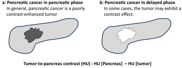
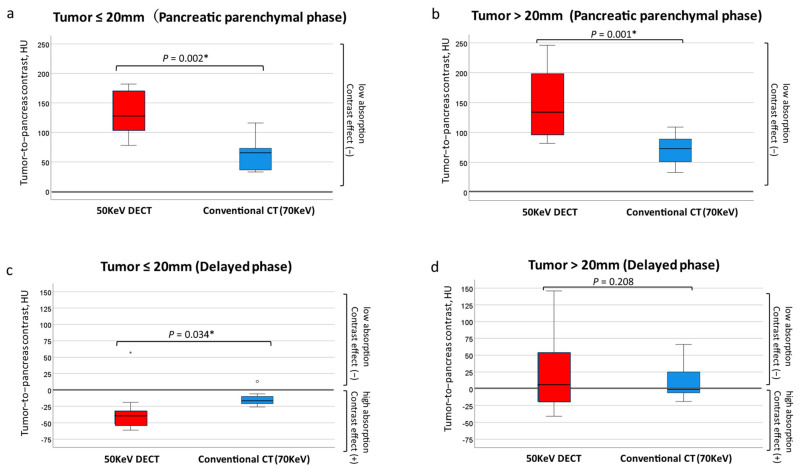
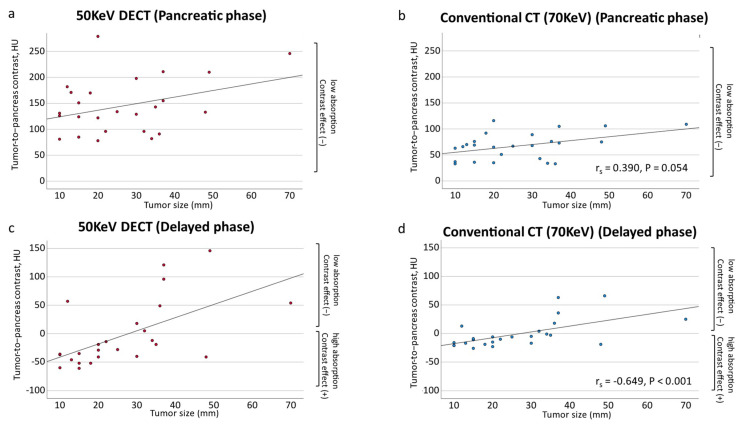
: The usefulness of dual-energy computed tomography (DECT) for low absorption in the parenchymal phase and contrast effects in the delayed phase for pancreatic cancer is not clear. Therefore, the diagnostic capability of low-KeV images obtained using DECT for pancreatic cancer in the pancreatic parenchymal and delayed phases was evaluated quantitatively and qualitatively. : Twenty-five patients with pancreatic cancer who underwent contrast-enhanced DECT were included. A total of 50 and 70 KeV CT images, classified as low-keV and conventional CT-equivalent images, were produced, respectively. The tumor-to-pancreas contrast (Hounsfield units [HU]) in the pancreatic parenchymal and delayed phases was calculated by subtracting the CT value of the pancreatic tumor from that of normal parenchyma. : The median tumor-to-pancreas contrast on 50 KeV CT in the pancreatic parenchymal phase (133 HU) was higher than that on conventional CT (68 HU) ( < 0.001). The median tumor-to-pancreas contrast in the delayed phase was -28 HU for 50 KeV CT and -9 HU for conventional CT ( = 0.545). For tumors < 20 mm, the tumor-to-pancreas contrast of 50 KeV CT (-39 HU) had a significantly clearer contrast effect than that of conventional CT (-16.5 HU), even in the delayed phase ( = 0.034). : These 50 KeV CT images may clarify the low-absorption areas of pancreatic cancer in the pancreatic parenchymal phase. A good contrast effect was observed in small pancreatic cancers on 50 KeV delayed-phase images, suggesting that DECT is useful for the visualization of early pancreatic cancer with a small tumor diameter.

摘要

双能 CT(DECT)在实质期低吸收率和延迟期对比效果方面对胰腺癌的作用尚不清楚。因此,本研究定量和定性评估了 DECT 低 keV 图像在胰腺实质期和延迟期对胰腺癌的诊断能力。

本研究纳入 25 例经增强 DECT 检查的胰腺癌患者。共生成 50 keV 和 70 keV CT 图像,分别归类为低 keV 和常规 CT 等效图像。通过从正常胰腺实质的 CT 值中减去胰腺肿瘤的 CT 值,计算胰腺实质期和延迟期肿瘤与胰腺的对比度(HU)。

50 keV CT 在胰腺实质期的肿瘤与胰腺对比度的中位数(133 HU)高于常规 CT(68 HU)( < 0.001)。50 keV CT 在延迟期的肿瘤与胰腺对比度为-28 HU,而常规 CT 为-9 HU( = 0.545)。对于 < 20 mm 的肿瘤,50 keV CT 的肿瘤与胰腺对比度(-39 HU)的对比度效果明显优于常规 CT(-16.5 HU),甚至在延迟期也是如此( = 0.034)。

这些 50 keV CT 图像可能会使胰腺癌在胰腺实质期的低吸收率区域更清晰。在 50 keV 延迟期图像上,较小的胰腺癌观察到良好的对比度效果,表明 DECT 对显示小肿瘤直径的早期胰腺癌有用。

https://cdn.ncbi.nlm.nih.gov/pmc/blobs/b6f0/11510840/5fe254aaeb19/tomography-10-00117-g006.jpg
https://cdn.ncbi.nlm.nih.gov/pmc/blobs/b6f0/11510840/25d63ff95225/tomography-10-00117-g001.jpg
https://cdn.ncbi.nlm.nih.gov/pmc/blobs/b6f0/11510840/1fac267caefd/tomography-10-00117-g002.jpg
https://cdn.ncbi.nlm.nih.gov/pmc/blobs/b6f0/11510840/9f1f81074320/tomography-10-00117-g003.jpg
https://cdn.ncbi.nlm.nih.gov/pmc/blobs/b6f0/11510840/6a80446d6c35/tomography-10-00117-g004.jpg
https://cdn.ncbi.nlm.nih.gov/pmc/blobs/b6f0/11510840/b1c95980a422/tomography-10-00117-g005.jpg
https://cdn.ncbi.nlm.nih.gov/pmc/blobs/b6f0/11510840/5fe254aaeb19/tomography-10-00117-g006.jpg

相似文献

1
Diagnostic Value of Contrast-Enhanced Dual-Energy Computed Tomography in the Pancreatic Parenchymal and Delayed Phases for Pancreatic Cancer.增强双能 CT 胰腺实质期和延迟期扫描对胰腺癌的诊断价值。
Tomography. 2024 Oct 7;10(10):1591-1604. doi: 10.3390/tomography10100117.
2
Optimized versus conventional trigger threshold for pancreatic phase image acquisition using dual-energy CT at 40-keV: a randomized controlled trial.40keV双能量CT胰腺期图像采集的优化触发阈值与传统触发阈值对比:一项随机对照试验
Abdom Radiol (NY). 2025 May;50(5):2304-2311. doi: 10.1007/s00261-024-04637-6. Epub 2024 Oct 21.
3
High-Pitch Multienergy Coronary CT Angiography in Dual-Source Photon-Counting Detector CT Scanner at Low Iodinated Contrast Dose.双源光子计数探测器 CT 扫描仪低碘对比剂剂量下的高频率多能量冠状动脉 CT 血管成像。
Invest Radiol. 2023 Sep 1;58(9):681-690. doi: 10.1097/RLI.0000000000000961.
4
Optimal virtual monoenergy for the detection of pancreatic adenocarcinoma during the pancreatic parenchymal phase on photon counting CT.光子计数CT胰腺实质期检测胰腺腺癌的最佳虚拟单能量
Abdom Radiol (NY). 2025 Jan 7. doi: 10.1007/s00261-024-04696-9.
5
Dual-energy CT Radiomics Combined with Quantitative Parameters for Differentiating Lung Adenocarcinoma From Squamous Cell Carcinoma: A Dual-center Study.双能量CT影像组学联合定量参数鉴别肺腺癌与肺鳞癌:一项双中心研究
Acad Radiol. 2025 Mar;32(3):1675-1684. doi: 10.1016/j.acra.2024.09.024. Epub 2024 Sep 25.
6
Comparing fully automated AI body composition biomarkers at differing virtual monoenergetic levels using dual-energy CT.使用双能CT比较不同虚拟单能量水平下的全自动人工智能身体成分生物标志物。
Abdom Radiol (NY). 2025 Jun;50(6):2758-2769. doi: 10.1007/s00261-024-04733-7. Epub 2024 Dec 7.
7
A Simplex algorithm approach to determine the optimal keV and window setting for appendicitis visualization.一种用于确定阑尾炎可视化的最佳keV和窗宽设置的单纯形算法方法。
Eur J Radiol. 2025 Oct;191:112282. doi: 10.1016/j.ejrad.2025.112282. Epub 2025 Jul 17.
8
Comparing two deep learning spectral reconstruction levels for abdominal evaluation using a rapid-kVp-switching dual-energy CT scanner.使用快速千伏切换双能CT扫描仪比较两种深度学习光谱重建水平用于腹部评估。
Abdom Radiol (NY). 2025 Mar 17. doi: 10.1007/s00261-025-04868-1.
9
Combining Low-energy Images in Dual-energy Spectral CT With Deep Learning Image Reconstruction Algorithm to Improve Inferior Vena Cava Image Quality.结合双能谱CT中的低能量图像与深度学习图像重建算法以提高下腔静脉图像质量
J Comput Assist Tomogr. 2025;49(4):604-610. doi: 10.1097/RCT.0000000000001713. Epub 2025 Jan 27.
10
Spectral CT imaging for assessment of metastases in melanoma patients: multi-reader evaluation.用于评估黑色素瘤患者转移灶的光谱CT成像:多阅片者评估
Cancer Imaging. 2025 Jun 16;25(1):74. doi: 10.1186/s40644-025-00889-7.

本文引用的文献

1
Uncommon Presentation of Gastric Duplication Cyst with Left-Sided Portal Hypertension: A Case Report and Literature Review.胃重复囊肿伴左侧门静脉高压的罕见表现:一例报告及文献复习
Diagnostics (Basel). 2024 Mar 22;14(7):675. doi: 10.3390/diagnostics14070675.
2
Early features of pancreatic cancer on magnetic resonance imaging (MRI): a case-control study.胰腺癌的磁共振成像(MRI)早期特征:一项病例对照研究。
Abdom Radiol (NY). 2024 May;49(5):1489-1501. doi: 10.1007/s00261-024-04271-2. Epub 2024 Apr 5.
3
Perineural invasion detection in pancreatic ductal adenocarcinoma using artificial intelligence.
使用人工智能检测胰腺导管腺癌的神经周围侵犯。
Sci Rep. 2023 Aug 21;13(1):13628. doi: 10.1038/s41598-023-40833-y.
4
Dual-energy CT of the pancreas: comparison between virtual non-contrast images and true non-contrast images in the detection of pancreatic lesion.胰腺双能量CT:虚拟平扫图像与真实平扫图像在胰腺病变检测中的比较
Abdom Radiol (NY). 2023 Aug;48(8):2596-2603. doi: 10.1007/s00261-023-03914-0. Epub 2023 May 20.
5
Value of Dual-Energy Computed Tomography for Detecting Small Pancreatic Ductal Adenocarcinoma.双能计算机断层扫描在检测小胰腺癌中的价值
Pancreas. 2022;51(10):1352-1358. doi: 10.1097/MPA.0000000000002207.
6
MRI vs. CT for pancreatic adenocarcinoma vascular invasion: comparative diagnostic test accuracy systematic review and meta-analysis.MRI 与 CT 对胰腺腺癌血管侵犯的诊断价值比较:系统评价和 Meta 分析。
Eur Radiol. 2023 Oct;33(10):6883-6891. doi: 10.1007/s00330-023-09659-0. Epub 2023 Apr 21.
7
Risk Assessment and Pancreatic Cancer: Diagnostic Management and Artificial Intelligence.风险评估与胰腺癌:诊断管理与人工智能
Cancers (Basel). 2023 Jan 5;15(2):351. doi: 10.3390/cancers15020351.
8
Comparison of image quality and pancreatic ductal adenocarcinoma conspicuity between the low-kVp and dual-energy CT reconstructed with deep-learning image reconstruction algorithm.低千伏与采用深度学习图像重建算法重建的双能CT之间图像质量及胰腺导管腺癌显示度的比较
Eur J Radiol. 2023 Feb;159:110685. doi: 10.1016/j.ejrad.2022.110685. Epub 2022 Dec 30.
9
Artificial intelligence using deep learning analysis of endoscopic ultrasonography images for the differential diagnosis of pancreatic masses.利用深度学习分析内镜超声图像的人工智能用于胰腺肿块的鉴别诊断。
Endoscopy. 2023 Feb;55(2):140-149. doi: 10.1055/a-1873-7920. Epub 2022 Jun 10.
10
Deep learning radiomics based on contrast-enhanced ultrasound images for assisted diagnosis of pancreatic ductal adenocarcinoma and chronic pancreatitis.基于对比增强超声图像的深度学习放射组学用于辅助诊断胰腺导管腺癌和慢性胰腺炎。
BMC Med. 2022 Mar 2;20(1):74. doi: 10.1186/s12916-022-02258-8.