Ríos-Anillo Margarita R, Ahmad Mostapha, Acosta-López Johan E, Cervantes-Henríquez Martha L, Henao-Castaño Maria C, Morales-Moreno Maria T, Espitia-Almeida Fabián, Vargas-Manotas José, Sánchez-Barros Cristian, Pineda David A, Sánchez-Rojas Manuel
Facultad de Ciencias de la Salud, Centro de Investigaciones en Ciencias de la Vida, Universidad Simón Bolívar, Barranquilla 080005, Colombia.
Médico Residente de Neurología, Facultad de Ciencias de la Salud, Centro de Investigaciones en Ciencias de la Vida, Universidad Simón Bolívar, Barranquilla 080005, Colombia.
Biomedicines. 2024 Sep 24;12(10):2166. doi: 10.3390/biomedicines12102166.
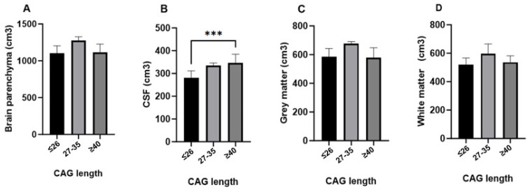
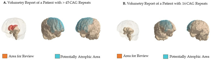
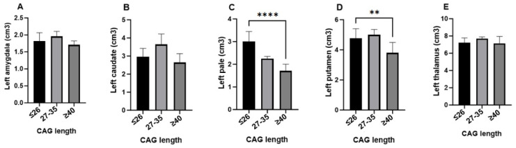
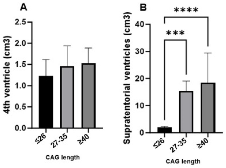
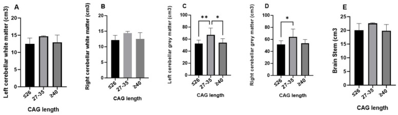
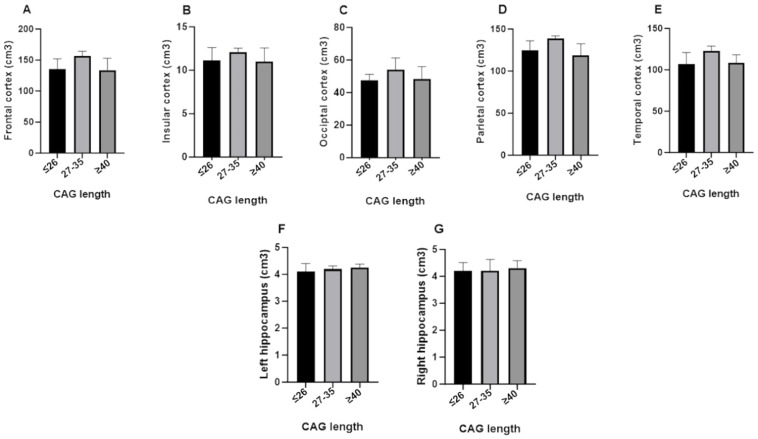
The premanifest phase of Huntington's disease (HD) is characterized by the absence of motor symptoms and exhibits structural changes in imaging that precede clinical manifestation. This study aimed to analyze volumetric changes identified through brain magnetic resonance imaging (MRI) processed using artificial intelligence (AI) software in premanifest HD individuals, focusing on the relationship between CAG triplet expansion and structural biomarkers. The study included 36 individuals descending from families affected by HD in the Department of Atlántico. Sociodemographic data were collected, followed by peripheral blood sampling to extract genomic DNA for quantifying CAG trinucleotide repeats in the Huntingtin gene. Brain volumes were evaluated using AI software (Entelai/IMEXHS, v4.3.4) based on MRI volumetric images. Correlations between brain volumes and variables such as age, sex, and disease status were determined. All analyses were conducted using SPSS (v. IBM SPSS Statistics 26), with significance set at < 0.05. The analysis of brain volumes according to CAG repeat expansion shows that individuals with ≥40 repeats evidence significant increases in cerebrospinal fluid (CSF) volume and subcortical structures such as the amygdalae and left caudate nucleus, along with marked reductions in cerebral white matter, the cerebellum, brainstem, and left pallidum. In contrast, those with <40 repeats show minimal or moderate volumetric changes, primarily in white matter and CSF. These findings suggest that CAG expansion selectively impacts key brain regions, potentially influencing the progression of Huntington's disease, and that AI in neuroimaging could identify structural biomarkers long before clinical symptoms appear.
亨廷顿舞蹈症(HD)的临床前期表现为无运动症状,且在影像学上呈现出早于临床表现的结构变化。本研究旨在分析使用人工智能(AI)软件处理的脑磁共振成像(MRI)在HD临床前期个体中识别出的体积变化,重点关注CAG三联体扩增与结构生物标志物之间的关系。该研究纳入了来自大西洋省受HD影响家庭的36名个体。收集了社会人口统计学数据数据数据数据,随后采集外周血样本以提取基因组DNA,用于定量亨廷顿基因中的CAG三核苷酸重复序列。基于MRI体积图像,使用AI软件(Entelai/IMEXHS,v4.3.4)评估脑容量。确定了脑容量与年龄、性别和疾病状态等变量之间的相关性。所有分析均使用SPSS(v. IBM SPSS Statistics 26)进行,显著性设定为<0.05。根据CAG重复扩增对脑容量进行的分析表明,CAG重复序列≥40次的个体脑脊液(CSF)体积以及杏仁核和左侧尾状核等皮质下结构显著增加,同时脑白质、小脑、脑干和左侧苍白球明显减少。相比之下,CAG重复序列<40次的个体体积变化最小或中等,主要发生在白质和脑脊液中。这些发现表明,CAG扩增选择性地影响关键脑区,可能影响亨廷顿舞蹈症的进展,并且神经影像学中的AI可以在临床症状出现之前很久就识别出结构生物标志物。