Tolstova Tatiana, Dotsenko Ekaterina, Luzgina Natalia, Rusanov Alexander
Institute of Biomedical Chemistry, Pogodinskaya 10, 119121 Moscow, Russia.
Biomedicines. 2024 Oct 2;12(10):2243. doi: 10.3390/biomedicines12102243.
Alzheimer's disease (AD) develops as a result of oxidative damage to neurons and chronic inflammation of microglia. These processes can be influenced by the use of a conditioned medium (CM) derived from mesenchymal stem cells (MSCs). The CM contains a wide range of factors that have neurotrophic, antioxidant, and anti-inflammatory effects. In addition, the therapeutic potential of the CM can be further enhanced by pretreating the MSCs to increase their paracrine activity. The current study aimed to investigate the neuroprotective effects of CM derived from MSCs, which were either activated by a TLR3 ligand or exposed to CoCl, a hypoxia mimetic (pCM or hCM, respectively), in an in vitro model of AD.
We have developed a novel in vitro model of AD that allows us to investigate the neuroprotective and anti-inflammatory effects of MSCs on induced neurodegeneration in the PC12 cell line and the activation of microglia using THP-1 cells.
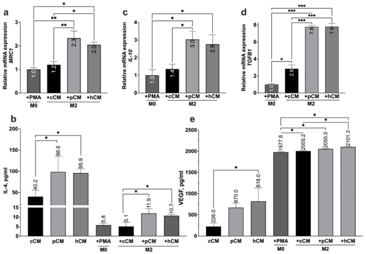
This study demonstrates for the first time that pCM and hCM exhibit more pronounced immunosuppressive effects on proinflammatory M1 macrophages compared to CM derived from untreated MSCs (cCM). This may help prevent the development of neuroinflammation by balancing the M1 and M2 microglial phenotypes via the decreased secretion of proinflammatory cytokines (IL-1β, IL-6, and TNF-α) and increased secretion of IL-4, as well as the expression of and by macrophages. Moreover, a previously unknown increase in the neurotrophic properties of hCM was discovered, which led to an increase in the viability of neuron-like PC12 cells under HO-induced oxidative-stress conditions. These results are likely associated with an increase in the production of growth factors, including vascular endothelial growth factor (VEGF). In addition, the neuroprotective effects of CM from preconditioned MSCs are also mediated by the activation of the Nrf2/ARE pathway in PC12 cells.
TLR3 activation in MSCs leads to more potent immunosuppressive effects of the CM against pro-inflammatory M1 macrophages, while the use of hCM led to increased neurotrophic effects after HO-induced damage to neuronal cells. These results are of interest for the potential treatment of AD with CM from preactivated MSCs.
阿尔茨海默病(AD)是神经元氧化损伤和小胶质细胞慢性炎症的结果。这些过程可能受到源自间充质干细胞(MSCs)的条件培养基(CM)的使用影响。该条件培养基含有多种具有神经营养、抗氧化和抗炎作用的因子。此外,通过预处理间充质干细胞以增加其旁分泌活性,可以进一步增强条件培养基的治疗潜力。本研究旨在探讨在AD体外模型中,由Toll样受体3(TLR3)配体激活的间充质干细胞或暴露于低氧模拟物氯化钴(分别为pCM或hCM)所产生的条件培养基对神经的保护作用。
我们建立了一种新型的AD体外模型,该模型使我们能够研究间充质干细胞对PC12细胞系中诱导的神经变性的神经保护和抗炎作用,以及使用THP-1细胞对小胶质细胞激活的影响。
本研究首次证明,与未处理的间充质干细胞(cCM)产生的条件培养基相比,pCM和hCM对促炎性M1巨噬细胞表现出更明显的免疫抑制作用。这可能有助于通过降低促炎细胞因子(IL-1β、IL-6和TNF-α)的分泌以及增加IL-4的分泌,以及巨噬细胞中 和 的表达,来平衡M1和M2小胶质细胞表型,从而预防神经炎症的发展。此外,还发现hCM的神经营养特性有此前未知的增加,这导致在HO诱导的氧化应激条件下,类神经元PC12细胞的活力增加。这些结果可能与包括血管内皮生长因子(VEGF)在内的生长因子产生增加有关。此外,预处理的间充质干细胞产生的条件培养基的神经保护作用也通过PC12细胞中Nrf2/ARE途径的激活来介导。
间充质干细胞中的TLR3激活导致条件培养基对促炎性M1巨噬细胞具有更强的免疫抑制作用,而使用hCM导致HO诱导的神经元细胞损伤后神经营养作用增强。这些结果对于用预激活的间充质干细胞产生的条件培养基潜在治疗AD具有重要意义。