Department of Emergency and Chest Pain Center, Qilu Hospital of Shandong University, Jinan, 250012, Shandong, People's Republic of China.
Shandong Provincial Clinical Research Center for Emergency and Critical Care Medicine, Institute of Emergency and Critical Care Medicine of Shandong University, Qilu Hospital of Shandong University, Jinan, 250012, Shandong, People's Republic of China.
Cardiovasc Diabetol. 2024 Oct 26;23(1):380. doi: 10.1186/s12933-024-02477-8.
Sodium-glucose cotransporter-2 inhibitors (SGLT2i) are now recommended for patients with heart failure, but the mechanisms that underlie the protective role of SGLT2i in cardiac remodeling remain unclear. Aldehyde dehydrogenase 2 (ALDH2) effectively prevents cardiac remodeling. Here, the key role of ALDH2 in the efficacy of SGLT2i on cardiac remodeling was studied.
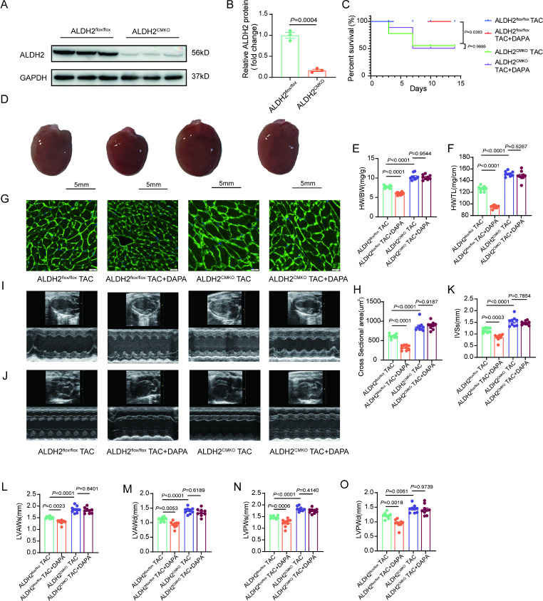
Analysis of multiple transcriptomic datasets and two-sample Mendelian randomization were performed to find out the differentially expressed genes between pathological cardiac hypertrophy models (patients) and controls. A pathological cardiac hypertrophy mouse model was established via transverse aortic constriction (TAC) or isoproterenol (ISO). Cardiomyocyte-specific ALDH2 knockout mice (ALDH2) and littermate control mice (ALDH2) were generated to determine the critical role of ALDH2 in the preventive effects of dapagliflozin (DAPA) on cardiac remodeling. RNA sequencing, gene knockdown or overexpression, bisulfite sequencing PCR, and luciferase reporter assays were performed to explore the underlying molecular mechanisms involved.
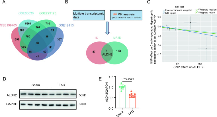
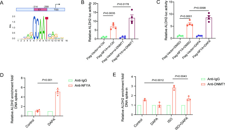
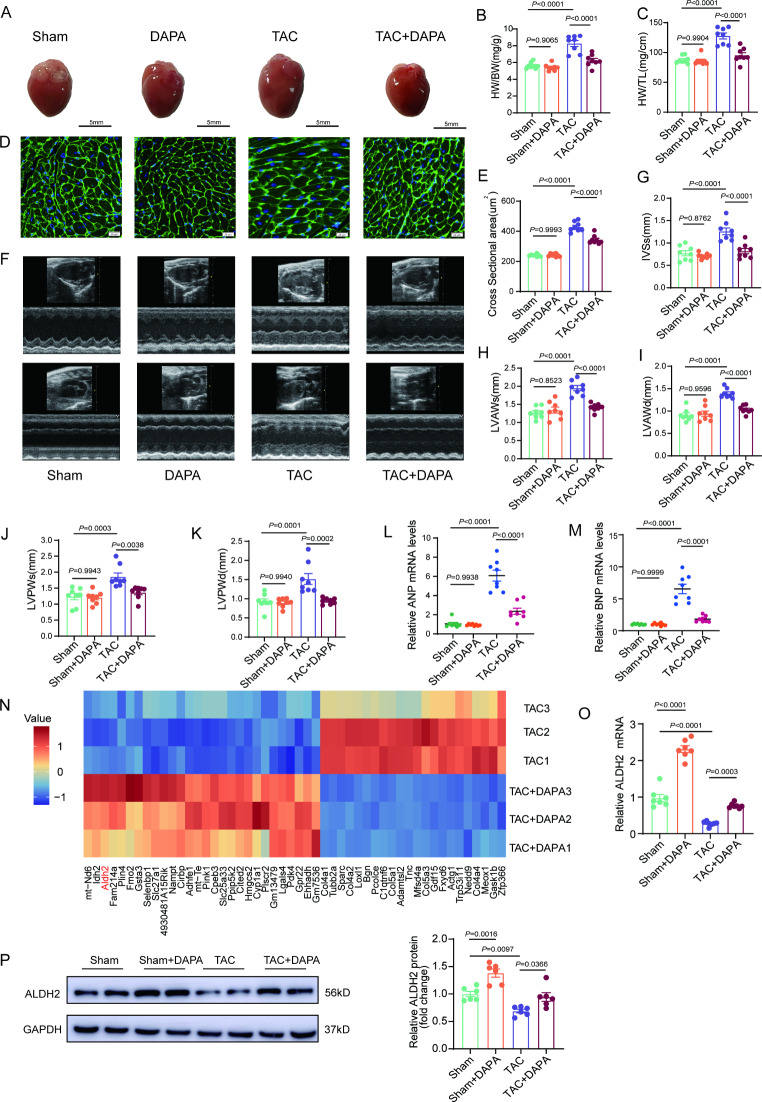
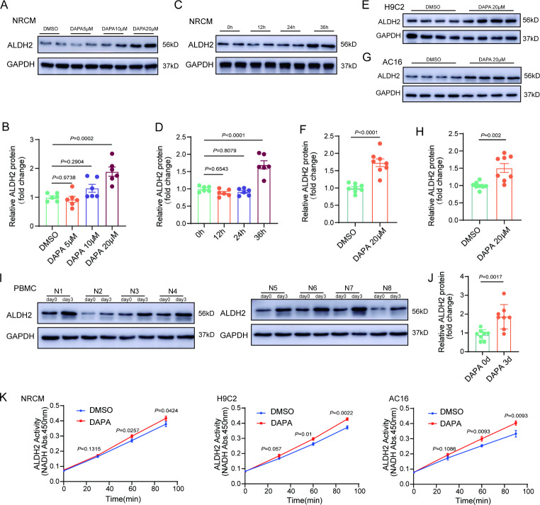
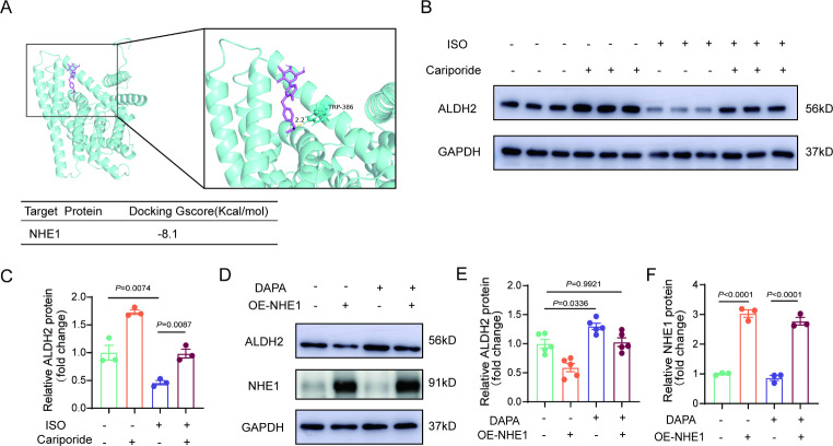
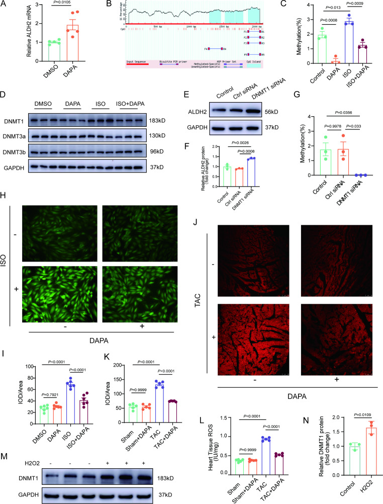
Only ALDH2 was differentially expressed when the differentially expressed genes obtained via Mendelian analysis and the differentially expressed genes obtained from the multiple transcriptome datasets were combined. Mendelian analysis revealed that ALDH2 was negatively related to the severity of myocardial hypertrophy in patients. DAPA alleviated cardiac remodeling in mouse hearts subjected to TAC or ISO. ALDH2 expression was reduced, whereas ALDH2 expression was restored by DAPA in hypertrophic hearts. Cardiomyocyte specific ALDH2 knockout abolished the protective role of DAPA in preventing cardiac remodeling. ALDH2 expression and activity were increased in DAPA-treated neonatal rat primary cardiomyocytes (NRCMs), H9C2 cells and AC16 cells. Moreover, DAPA upregulated ALDH2 in peripheral blood mononuclear cells (PBMCs) from patients with type 2 diabetes. Sodium/proton exchanger 1 (NHE1) inhibition contributed to the regulation of ALDH2 by DAPA. DAPA suppressed the production of reactive oxygen species (ROS), downregulated DNA methyltransferase 1 (DNMT1) and subsequently reduced the ALDH2 promoter methylation level. Further studies revealed that DAPA enhanced the binding of nuclear transcription factor Y, subunit A (NFYA) to the promoter region of ALDH2, which was due to the decreased promoter methylation level of ALDH2.
The upregulation of ALDH2 plays a critical role in the protection of DAPA against cardiac remodeling. DAPA enhances the binding of NFYA to the ALDH2 promoter by reducing the ALDH2 promoter methylation level through NHE1/ROS/DNMT1 pathway.
钠-葡萄糖共转运蛋白 2 抑制剂(SGLT2i)现被推荐用于心力衰竭患者,但 SGLT2i 在心脏重构中发挥保护作用的机制仍不清楚。醛脱氢酶 2(ALDH2)可有效预防心脏重构。本研究旨在探讨 ALDH2 在 SGLT2i 治疗心脏重构疗效中的关键作用。
分析多个转录组数据集并进行两样本 Mendelian 随机化,以找出病理性心肌肥厚模型(患者)与对照之间差异表达的基因。通过主动脉缩窄(TAC)或异丙肾上腺素(ISO)建立病理性心肌肥厚小鼠模型。生成心肌细胞特异性 ALDH2 敲除小鼠(ALDH2)和同窝对照小鼠(ALDH2),以确定 ALDH2 在达格列净(DAPA)预防心脏重构中的关键作用。进行 RNA 测序、基因敲低或过表达、亚硫酸氢盐测序 PCR 和荧光素酶报告基因检测,以探讨相关的分子机制。
仅当将 Mendelian 分析获得的差异表达基因与多个转录组数据集获得的差异表达基因相结合时,才发现 ALDH2 是差异表达的。Mendelian 分析表明,ALDH2 与患者心肌肥厚的严重程度呈负相关。DAPA 减轻了 TAC 或 ISO 处理的小鼠心脏的重构。在肥厚的心脏中,ALDH2 的表达减少,但 DAPA 可恢复其表达。心肌细胞特异性 ALDH2 敲除消除了 DAPA 预防心脏重构的保护作用。在 DAPA 处理的新生大鼠原代心肌细胞(NRCMs)、H9C2 细胞和 AC16 细胞中,ALDH2 的表达和活性增加。此外,DAPA 可增加 2 型糖尿病患者外周血单个核细胞(PBMCs)中的 ALDH2 表达。钠/质子交换体 1(NHE1)抑制作用有助于 DAPA 对 ALDH2 的调节。DAPA 抑制活性氧(ROS)的产生,下调 DNA 甲基转移酶 1(DNMT1),从而降低 ALDH2 启动子的甲基化水平。进一步的研究表明,DAPA 通过降低 ALDH2 启动子的甲基化水平,增强核转录因子 Y,亚基 A(NFYA)与 ALDH2 启动子区域的结合,这归因于 ALDH2 启动子的甲基化水平降低。
ALDH2 的上调在 DAPA 对抗心脏重构的保护中起着关键作用。DAPA 通过 NHE1/ROS/DNMT1 途径降低 ALDH2 启动子的甲基化水平,从而增强 NFYA 与 ALDH2 启动子的结合。