Dementia Research Centre, UCL Queen Square Institute of Neurology, University College London, London, UK.
Dementia Research Institute, University College London, London, UK.
J Neurol Neurosurg Psychiatry. 2024 Jul 15;95(8):748-752. doi: 10.1136/jnnp-2023-332067.
Consistent patterns of reduced cortical thickness have been identified in early Alzheimer's disease (AD). However, the pathological factors that influence rates of cortical thinning within these AD signature regions remain unclear.
Participants were from the Insight 46 substudy of the MRC National Survey of Health and Development (NSHD; 1946 British birth cohort), a prospective longitudinal cohort study. Linear regression was used to examine associations of baseline cerebral β-amyloid (Aβ) deposition, measured using florbetapir positron emission tomography, and baseline white matter hyperintensity volume (WMHV) on MRI, a marker of cerebral small vessel disease, with subsequent longitudinal changes in AD signature cortical thickness quantified from baseline and repeat MRI (mean [SD] interval 2.4 [0.2] years).
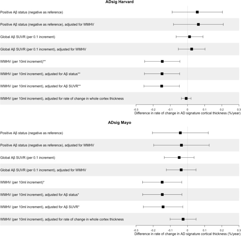
In a population-based sample of 337 cognitively normal older white adults (mean [SD] age at baseline 70.5 [0.6] years; 48.1% female), higher global WMHV at baseline related to faster subsequent rates of cortical thinning in both AD signature regions (~0.15%/year faster per 10 mL additional WMHV), whereas baseline Aβ status did not. Among Aβ positive participants (n=56), there was some evidence that greater global Aβ standardised uptake value ratio at baseline related to faster cortical thinning in the AD signature Mayo region, but this did not reach statistical significance (p=0.08).
Cortical thinning within AD signature regions may develop via cerebrovascular pathways. Perhaps reflecting the age of the cohort and relatively low prevalence of Aβ-positivity, robust Aβ-related differences were not detected. Longitudinal follow-up incorporating additional biomarkers will allow assessment of how these relationships evolve closer to expected dementia onset.
在早期阿尔茨海默病(AD)中,已经确定了皮质厚度减少的一致模式。然而,影响这些 AD 特征区域皮质变薄速度的病理因素尚不清楚。
参与者来自 MRC 国家健康与发展调查(NSHD;1946 年英国出生队列)Insight 46 子研究,这是一项前瞻性纵向队列研究。线性回归用于检查基线脑β-淀粉样蛋白(Aβ)沉积(使用 florbetapir 正电子发射断层扫描测量)与基线磁共振成像(MRI)上的白质高信号体积(WMHV)(脑小血管疾病的标志物)与 AD 特征皮质厚度的后续纵向变化之间的关联,AD 特征皮质厚度是从基线和重复 MRI 中量化得出的(平均[标准差]间隔 2.4[0.2]年)。
在基于人群的 337 名认知正常的老年白人成年人样本中(基线时的平均[标准差]年龄为 70.5[0.6]岁;48.1%为女性),基线时较高的全球 WMHV 与 AD 特征区域中更快的皮质变薄速度有关(每增加 10 mL 额外的 WMHV 就会增加约 0.15%/年),而基线 Aβ 状态则没有。在 Aβ 阳性参与者中(n=56),有一些证据表明,基线时较大的全球 Aβ 标准化摄取比值与 AD 特征 Mayo 区域更快的皮质变薄有关,但这并未达到统计学意义(p=0.08)。
AD 特征区域内的皮质变薄可能通过脑血管途径发展。也许反映了队列的年龄和相对较低的 Aβ 阳性率,没有检测到与 Aβ 相关的显著差异。纳入额外生物标志物的纵向随访将能够评估这些关系在接近预期痴呆发作时的演变情况。