Goris Liselot C, Mikerov Mikhail, Pautasso Juan J, Sechopoulos Ioannis
Technical Medicine Centre, University of Twente, Enschede, The Netherlands.
Department of Medical Imaging, Radboud University Medical Center, Nijmegen, The Netherlands.
Med Phys. 2025 Feb;52(2):1037-1044. doi: 10.1002/mp.17497. Epub 2024 Oct 29.
In 4D dynamic contrast-enhanced dedicated breast computed tomography (4D DCE-bCT), the functional properties of the breast will be characterized by monitoring the uptake and washout of iodine-based contrast agents over time. This information could be valuable in breast cancer treatment. However, prior to clinical implementation, it is crucial to validate the quantitative estimates of iodine concentrations at each time point during acquisition.
To develop an in-line spectroscopy system capable of measuring iodine concentrations in a dynamic x-ray breast phantom in real-time.
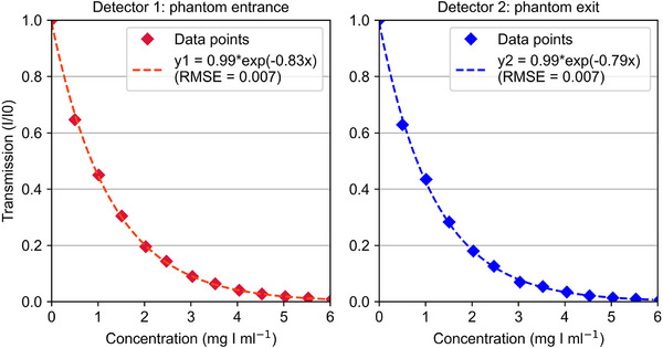
Potassium iodide served as the contrast agent. The system was set-up at both the entrance and exit of the phantom. It comprises a fiber-coupled green LED and collimator, which together ensure that a parallel beam passes through the sample holder. Transmitted light is captured by a collimator on the opposite side and directed through a fiber optic cable to a photodetector for intensity measurement. The relationship between 13 iodine concentrations (0-6 mg I/mL) and light transmission was tested, and the system's repeatability and accuracy were determined.
The system exhibited a strong correlation between iodine concentration and transmission values, achieving a root-mean-square error of 0.007. The repeated measurements had relative standard deviations of 0.04% and 0.1% for repeated water measurements at the phantom's entrance and exit, respectively. Furthermore, the accuracy measurements gave a mean error of fitting of 0.008 (± 0.07) mg I/mL.
The in-line spectroscopy system can effectively monitor iodine concentrations in a dynamic breast phantom, providing a reliable method for quantitative validation of 4D DCE-bCT.
在四维动态对比增强专用乳腺计算机断层扫描(4D DCE-bCT)中,通过监测碘基造影剂随时间的摄取和洗脱来表征乳腺的功能特性。这些信息在乳腺癌治疗中可能具有重要价值。然而,在临床应用之前,验证采集过程中每个时间点碘浓度的定量估计至关重要。
开发一种能够实时测量动态乳腺X线体模中碘浓度的在线光谱系统。
碘化钾用作造影剂。该系统设置在体模的入口和出口处。它包括一个光纤耦合的绿色发光二极管和准直器,共同确保平行光束穿过样品架。透射光由另一侧的准直器捕获,并通过光纤电缆导向光电探测器进行强度测量。测试了13种碘浓度(0-6 mg I/mL)与透光率之间的关系,并确定了系统的重复性和准确性。
该系统在碘浓度与透射值之间表现出很强的相关性,均方根误差为0.007。在体模入口和出口处对水进行重复测量时,重复测量的相对标准偏差分别为0.04%和0.1%。此外,准确性测量的拟合平均误差为0.008(±0.07)mg I/mL。
在线光谱系统能够有效地监测动态乳腺体模中的碘浓度,为4D DCE-bCT的定量验证提供了一种可靠的方法。