• 文献检索
  • 文档翻译
  • 深度研究
  • 学术资讯
  • Suppr Zotero 插件Zotero 插件
  • 邀请有礼
  • 套餐&价格
  • 历史记录
应用&插件
Suppr Zotero 插件Zotero 插件浏览器插件Mac 客户端Windows 客户端微信小程序
定价
高级版会员购买积分包购买API积分包
服务
文献检索文档翻译深度研究API 文档MCP 服务
关于我们
关于 Suppr公司介绍联系我们用户协议隐私条款
关注我们

Suppr 超能文献

核心技术专利:CN118964589B侵权必究
粤ICP备2023148730 号-1Suppr @ 2026

文献检索

告别复杂PubMed语法,用中文像聊天一样搜索,搜遍4000万医学文献。AI智能推荐,让科研检索更轻松。

立即免费搜索

文件翻译

保留排版,准确专业,支持PDF/Word/PPT等文件格式,支持 12+语言互译。

免费翻译文档

深度研究

AI帮你快速写综述,25分钟生成高质量综述,智能提取关键信息,辅助科研写作。

立即免费体验

在X连锁视网膜劈裂症小鼠模型中,一种渗透压依赖性机制可部分改善视网膜囊肿并挽救视锥细胞功能。

An osmolarity dependent mechanism partially ameliorates retinal cysts and rescues cone function in a mouse model of X-linked retinoschisis.

作者信息

Gehrke Ella J, Thompson Jacob, Kalmanek Emily, Stanley Sarah T, Laird Joseph, Bhattarai Sajag, Lobeck Brianna, Mayer Sara, Mahoney Angela, Hassan Salma, Hsu Ying, Drack Arlene

机构信息

Department of Ophthalmology and Visual Sciences, Institute for Vision Research, University of Iowa, Iowa City, IA, United States.

Department of Epidemiology, College of Public Health, University of Iowa, Iowa City, IA, United States.

出版信息

Front Med (Lausanne). 2024 Oct 15;11:1302119. doi: 10.3389/fmed.2024.1302119. eCollection 2024.

DOI:10.3389/fmed.2024.1302119
PMID:39473494
原文链接:https://pmc.ncbi.nlm.nih.gov/articles/PMC11518720/
Abstract

INTRODUCTION

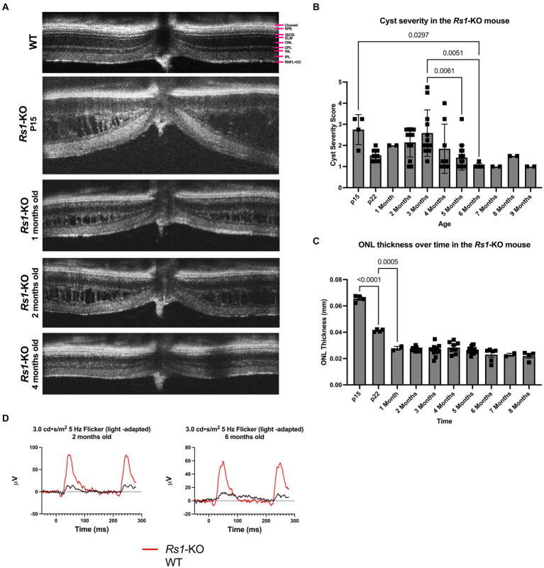
X-linked retinoschisis (XLRS) is a vitreoretinal dystrophy caused by gene mutations which disrupt retinoschisin-1 (RS1) function. Vital for retinal architecture, the absence of functional RS1 leads to the development of intraretinal cysts. Intravitreal injection of a gene therapy for treating XLRS caused ocular inflammation in high dose groups in a phase I/II clinical trial. This study investigates a low dose subretinal gene therapy in knockout (-KO) mice compared to injection of buffer alone. Observation of an unexpected therapeutic effect following the subretinal injection of the hypertonic buffer led to novel findings in XLRS.

METHODS

-KO mice were subretinally injected with an AAV2/4 vector ( = 10) containing the gene driven by an Ef1α promoter, a hypertonic buffer ( = 15) (180 mM NaCl 0.001% F68/PBS (pH 7.4)), or isotonic buffer ( = 7) (155.2 mM NaCl 0.001% F68/PBS, pH 7.0). A sham puncture group was also included ( = 6). Endpoints included electroretinogram (ERG), optical coherence tomography (OCT), a visually guided swim assay (VGSA), and immunohistochemistry.

RESULTS

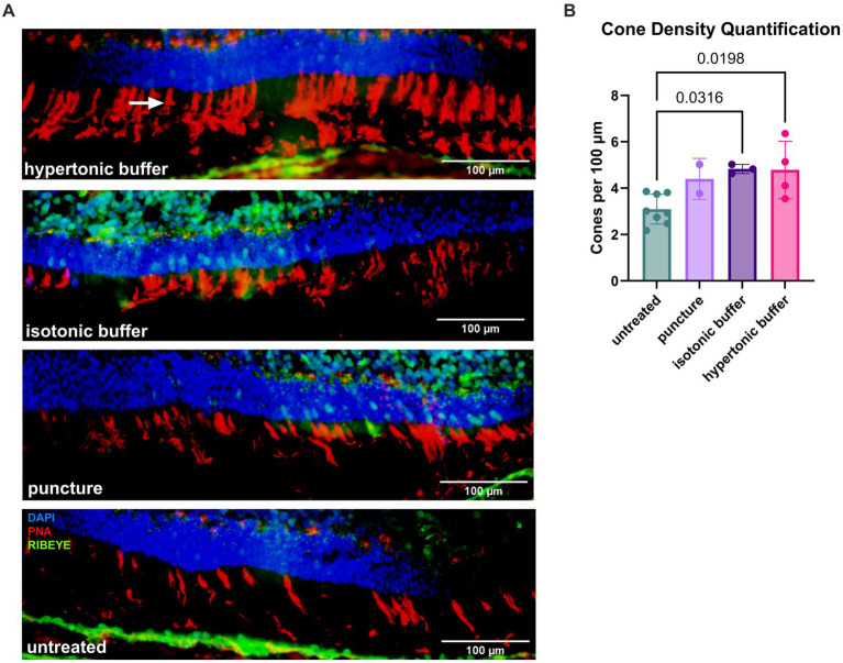
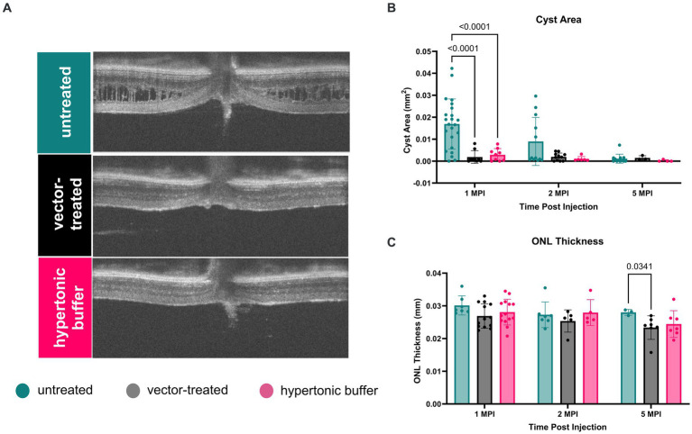
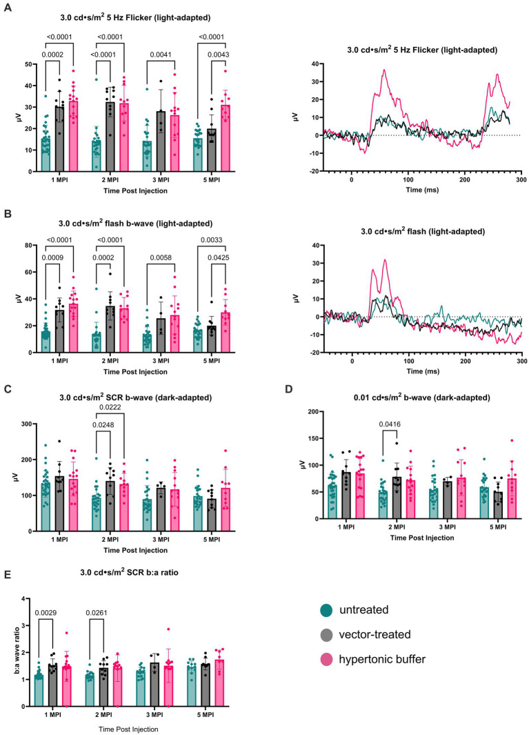
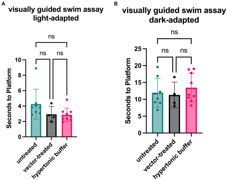
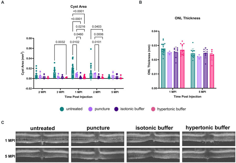
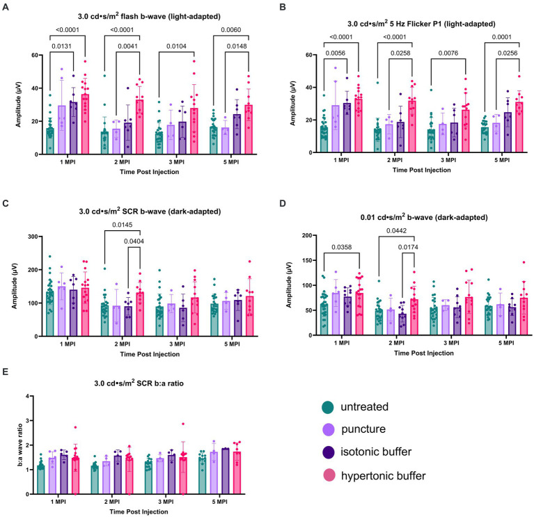
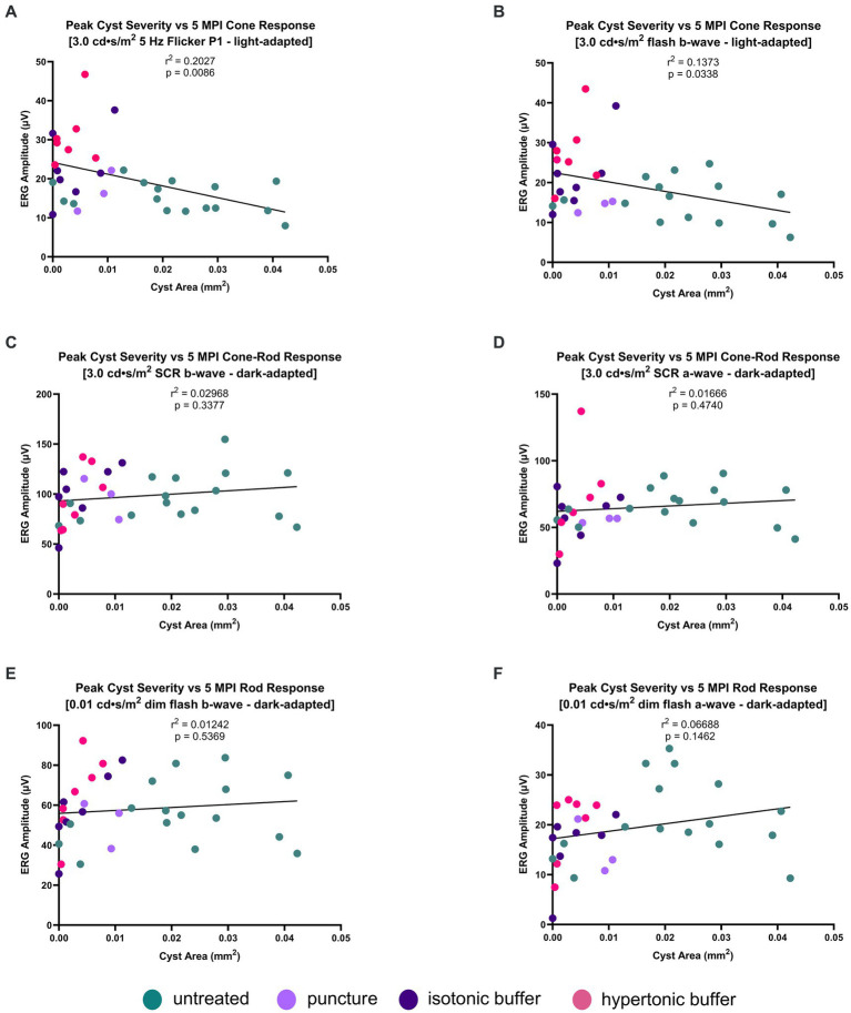
Unexpectedly, hypertonic buffer-injected eyes had reduced cyst severity at 1-month post-injection (MPI) ( < 0.0001), higher amplitudes in cone-dominant ERGs persisting to 5 MPI (5 Hz flicker;  < 0.0001; 3.0 flash;  = 0.0033) and a trend for improved navigational vision in the light compared to untreated -KO eyes. To investigate the role of tonicity on this effect, an isotonic buffer-injected cohort was created (155.2 mM NaCl 0.001% F68/PBS, pH 7.0) ( = 7). Surprisingly, hypertonic buffer-injected eyes exhibited a greater reduction in cyst severity and demonstrated improved cone-dominant ERG metrics over isotonic buffer-injected and sham puncture eyes. An immunohistochemistry assay demonstrated greater cone density in hypertonic buffer-injected eyes than untreated -KO eyes at 5-6 MPI ( = 0.0198), suggesting a possible cone preservation mechanism. Moreover, our findings reveal a negative correlation between the peak severity of cysts and long-term ERG amplitudes in cone-dominant pathways, implying that effectively managing cysts could yield enduring benefits for cone function.

DISCUSSION/CONCLUSION: This study presents evidence that cyst resolution can be triggered through an osmolarity-dependent pathway, and early cyst resolution has long-term effects on cone signaling and survival, offering potential insights for the development of novel treatments for XLRS patients.

摘要

引言

X连锁视网膜劈裂症(XLRS)是一种由基因突变导致视网膜劈裂蛋白-1(RS1)功能破坏引起的玻璃体视网膜营养不良。RS1对视网膜结构至关重要,缺乏功能性RS1会导致视网膜内囊肿的形成。在一项I/II期临床试验中,高剂量组玻璃体内注射治疗XLRS的基因疗法引发了眼部炎症。本研究将低剂量视网膜下基因疗法与单独注射缓冲液相比较,在基因敲除(-KO)小鼠中进行研究。视网膜下注射高渗缓冲液后观察到意外的治疗效果,从而在XLRS研究中获得了新发现。

方法

向-KO小鼠视网膜下注射含由Ef1α启动子驱动的RS1基因的AAV2/4载体(n = 10)、高渗缓冲液(n = 15)(180 mM NaCl 0.001% F68/PBS(pH 7.4))或等渗缓冲液(n = 7)(155.2 mM NaCl 0.001% F68/PBS,pH 7.0)。还纳入了假穿刺组(n = 6)。观察指标包括视网膜电图(ERG)、光学相干断层扫描(OCT)、视觉引导游泳试验(VGSA)和免疫组织化学。

结果

出乎意料的是,注射高渗缓冲液的眼睛在注射后1个月(MPI)时囊肿严重程度降低(P < 0.0001),在以视锥细胞为主的ERG中较高的振幅持续至5 MPI(5 Hz闪烁;P < 0.0001;3.0闪光;P = 0.0033),并且与未治疗的-KO眼睛相比,在明亮环境中有改善导航视觉的趋势。为了研究渗透压对这种效应的作用,创建了注射等渗缓冲液的队列(155.2 mM NaCl 0.001% F68/PBS,pH 7.0)(n = 7)。令人惊讶的是,与注射等渗缓冲液和假穿刺的眼睛相比,注射高渗缓冲液的眼睛囊肿严重程度降低更大,并且以视锥细胞为主的ERG指标得到改善。免疫组织化学分析表明,在5 - 6 MPI时,注射高渗缓冲液的眼睛视锥细胞密度高于未治疗的-KO眼睛(P = 0.0198),提示可能存在视锥细胞保护机制。此外,我们的研究结果揭示了囊肿峰值严重程度与以视锥细胞为主的通路中长期ERG振幅之间的负相关,这意味着有效控制囊肿可能对视锥细胞功能产生持久益处。

讨论/结论:本研究提供的证据表明,囊肿消退可通过渗透压依赖性途径触发,早期囊肿消退对视锥细胞信号传导和存活具有长期影响,为开发针对XLRS患者的新疗法提供了潜在的见解。

https://cdn.ncbi.nlm.nih.gov/pmc/blobs/0c57/11518720/0e4cf19bb716/fmed-11-1302119-g008.jpg
https://cdn.ncbi.nlm.nih.gov/pmc/blobs/0c57/11518720/9b865f307d1b/fmed-11-1302119-g001.jpg
https://cdn.ncbi.nlm.nih.gov/pmc/blobs/0c57/11518720/ca231585d605/fmed-11-1302119-g002.jpg
https://cdn.ncbi.nlm.nih.gov/pmc/blobs/0c57/11518720/cf621fe54fdb/fmed-11-1302119-g003.jpg
https://cdn.ncbi.nlm.nih.gov/pmc/blobs/0c57/11518720/778a831d0c8d/fmed-11-1302119-g004.jpg
https://cdn.ncbi.nlm.nih.gov/pmc/blobs/0c57/11518720/0444c8634d74/fmed-11-1302119-g005.jpg
https://cdn.ncbi.nlm.nih.gov/pmc/blobs/0c57/11518720/111730cda997/fmed-11-1302119-g006.jpg
https://cdn.ncbi.nlm.nih.gov/pmc/blobs/0c57/11518720/0c87487aa38e/fmed-11-1302119-g007.jpg
https://cdn.ncbi.nlm.nih.gov/pmc/blobs/0c57/11518720/0e4cf19bb716/fmed-11-1302119-g008.jpg

相似文献

1
An osmolarity dependent mechanism partially ameliorates retinal cysts and rescues cone function in a mouse model of X-linked retinoschisis.在X连锁视网膜劈裂症小鼠模型中,一种渗透压依赖性机制可部分改善视网膜囊肿并挽救视锥细胞功能。
Front Med (Lausanne). 2024 Oct 15;11:1302119. doi: 10.3389/fmed.2024.1302119. eCollection 2024.
2
The dose-response relationship of subretinal gene therapy with rAAV2tYF-CB-h in a mouse model of X-linked retinoschisis.在X连锁视网膜劈裂症小鼠模型中,采用rAAV2tYF-CB-h进行视网膜下基因治疗的剂量反应关系。
Front Med (Lausanne). 2024 Feb 13;11:1304819. doi: 10.3389/fmed.2024.1304819. eCollection 2024.
3
AAV2/4-RS1 gene therapy in the retinoschisin knockout mouse model of X-linked retinoschisis.AAV2/4-RS1 基因治疗在 X 连锁性视网膜劈裂症的视蛋白敲除小鼠模型中的应用。
PLoS One. 2022 Dec 7;17(12):e0276298. doi: 10.1371/journal.pone.0276298. eCollection 2022.
4
Investigating the role of Caspase-1 in a mouse model of Juvenile X-linked Retinoschisis.研究半胱天冬酶-1在青少年X连锁视网膜劈裂症小鼠模型中的作用。
Front Med (Lausanne). 2024 Jun 13;11:1347599. doi: 10.3389/fmed.2024.1347599. eCollection 2024.
5
Of men and mice: Human X-linked retinoschisis and fidelity in mouse modeling.关于人类与小鼠:人类X连锁视网膜劈裂症及小鼠建模的准确性
Prog Retin Eye Res. 2022 Mar;87:100999. doi: 10.1016/j.preteyeres.2021.100999. Epub 2021 Aug 11.
6
Preclinical Dose-Escalation Study of Intravitreal AAV-RS1 Gene Therapy in a Mouse Model of X-linked Retinoschisis: Dose-Dependent Expression and Improved Retinal Structure and Function.玻璃体腔内注射腺相关病毒-RS1基因疗法在X连锁视网膜劈裂小鼠模型中的临床前剂量递增研究:剂量依赖性表达及视网膜结构与功能改善
Hum Gene Ther. 2016 May;27(5):376-89. doi: 10.1089/hum.2015.142.
7
An ex vivo gene therapy approach in X-linked retinoschisis.X连锁视网膜劈裂症的一种离体基因治疗方法。
Mol Vis. 2016 Jun 24;22:718-33. eCollection 2016.
8
Targeted Expression of Retinoschisin by Retinal Bipolar Cells in XLRS Promotes Resolution of Retinoschisis Cysts Sans RS1 From Photoreceptors.视网膜双极细胞靶向表达视锥视蛋白可促进 XLRS 患者 RS1 缺失型格子样变性的裂孔闭合而不影响感光细胞。
Invest Ophthalmol Vis Sci. 2022 Oct 3;63(11):8. doi: 10.1167/iovs.63.11.8.
9
Synaptic pathology in retinoschisis knockout (Rs1-/y) mouse retina and modification by rAAV-Rs1 gene delivery.视网膜劈裂敲除(Rs1-/y)小鼠视网膜中的突触病理学及rAAV-Rs1基因递送的修饰作用
Invest Ophthalmol Vis Sci. 2008 Aug;49(8):3677-86. doi: 10.1167/iovs.07-1071.
10
A phenotype-genotype correlation study of X-linked retinoschisis.X 连锁性视网膜劈裂的表型-基因型相关性研究。
Ophthalmology. 2013 Jul;120(7):1454-64. doi: 10.1016/j.ophtha.2012.12.008. Epub 2013 Feb 28.

本文引用的文献

1
The dose-response relationship of subretinal gene therapy with rAAV2tYF-CB-h in a mouse model of X-linked retinoschisis.在X连锁视网膜劈裂症小鼠模型中,采用rAAV2tYF-CB-h进行视网膜下基因治疗的剂量反应关系。
Front Med (Lausanne). 2024 Feb 13;11:1304819. doi: 10.3389/fmed.2024.1304819. eCollection 2024.
2
Morphological and functional parameters in X-linked retinoschisis patients-A multicentre retrospective cohort study.X连锁视网膜劈裂症患者的形态学和功能参数——一项多中心回顾性队列研究
Front Med (Lausanne). 2024 Jan 11;10:1331889. doi: 10.3389/fmed.2023.1331889. eCollection 2023.
3
A visually guided swim assay for mouse models of human retinal disease recapitulates the multi-luminance mobility test in humans.
一种用于人类视网膜疾病小鼠模型的视觉引导游泳试验概括了人类的多亮度移动性测试。
Saudi J Ophthalmol. 2023 Nov 18;37(4):313-320. doi: 10.4103/sjopt.sjopt_155_23. eCollection 2023 Oct-Dec.
4
X-linked Retinoschisis with Near-Normal Macular Structure and Function.具有近乎正常黄斑结构和功能的X连锁视网膜劈裂症
Ophthalmol Retina. 2023 Oct;7(10):847. doi: 10.1016/j.oret.2023.05.003. Epub 2023 May 31.
5
Subretinal gene therapy delays vision loss in a Bardet-Biedl Syndrome type 10 mouse model.视网膜下基因治疗延缓了10型巴德-比德尔综合征小鼠模型的视力丧失。
Mol Ther Nucleic Acids. 2022 Dec 12;31:164-181. doi: 10.1016/j.omtn.2022.12.007. eCollection 2023 Mar 14.
6
AAV2/4-RS1 gene therapy in the retinoschisin knockout mouse model of X-linked retinoschisis.AAV2/4-RS1 基因治疗在 X 连锁性视网膜劈裂症的视蛋白敲除小鼠模型中的应用。
PLoS One. 2022 Dec 7;17(12):e0276298. doi: 10.1371/journal.pone.0276298. eCollection 2022.
7
Intravitreal Delivery of rAAV2tYF-CB-hRS1 Vector for Gene Augmentation Therapy in Patients with X-Linked Retinoschisis: 1-Year Clinical Results.眼内注射 rAAV2tYF-CB-hRS1 载体进行 X 连锁性视网膜劈裂症基因增强治疗的 1 年临床结果。
Ophthalmol Retina. 2022 Dec;6(12):1130-1144. doi: 10.1016/j.oret.2022.06.013. Epub 2022 Jun 30.
8
ISCEV Standard for full-field clinical electroretinography (2022 update).国际临床电生理学会标准:全视野临床视网膜电流图(2022 更新版)。
Doc Ophthalmol. 2022 Jun;144(3):165-177. doi: 10.1007/s10633-022-09872-0. Epub 2022 May 5.
9
Hypertonic saline improves brain edema resulting from traumatic brain injury by suppressing the NF-κB/IL-1β signaling pathway and AQP4.高渗盐水通过抑制NF-κB/IL-1β信号通路和水通道蛋白4(AQP4)来改善创伤性脑损伤所致的脑水肿。
Exp Ther Med. 2020 Nov;20(5):71. doi: 10.3892/etm.2020.9199. Epub 2020 Sep 9.
10
Efficacy of topical brinzolamide in children with retinal dystrophies.局部用布林佐胺治疗视网膜营养不良患儿的疗效。
Ophthalmic Genet. 2019 Aug;40(4):350-358. doi: 10.1080/13816810.2019.1660381. Epub 2019 Sep 9.