Luyken Adrian Konstantin, Lappe Chris, Viard Romain, Löhle Matthias, Kleinlein Hanna Rebekka, Kuchcinski Grégory, Langner Sönke, Wenzel Anne-Marie, Walter Michael, Weber Marc-André, Storch Alexander, Devos David, Walter Uwe
Department of Neurology, Rostock University Medical Center, Gehlsheimer Str. 20, 18147, Rostock, Germany.
Institute of Diagnostic and Interventional Radiology, Pediatric Radiology and Neuroradiology, University Medical Center Rostock, Rostock, Germany.
J Neural Transm (Vienna). 2025 Mar;132(3):407-417. doi: 10.1007/s00702-024-02856-1. Epub 2024 Nov 1.
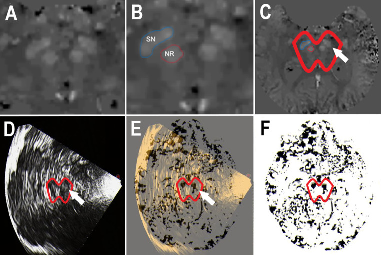
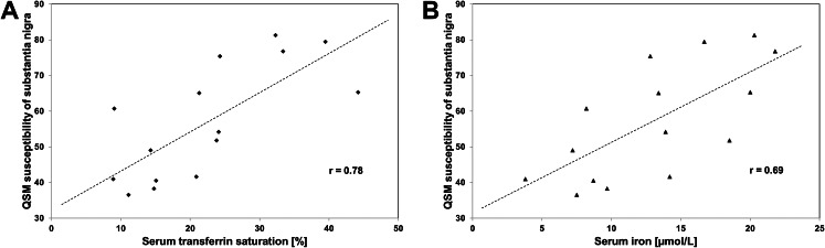
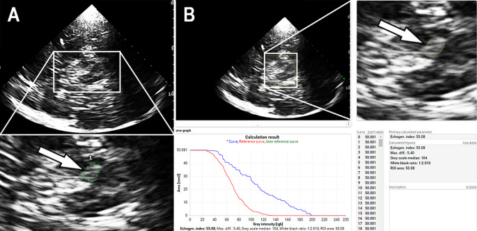
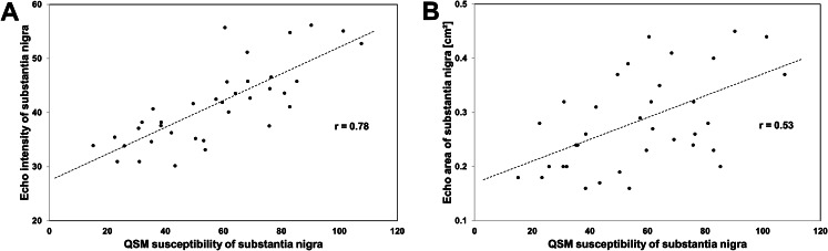
Quantitative susceptibility mapping (QSM) and transcranial sonography (TCS) offer proximal evaluations of iron load in the substantia nigra. Our prospective study aimed to investigate the relationship between QSM and TCS measurements of nigral iron content in patients with Parkinson's disease (PD). In secondary analyses, we wanted to explore the correlation of substantia nigra imaging data with clinical and laboratory findings. Eighteen magnetic resonance imaging and TCS examinations were performed in 15 PD patients at various disease stages. Susceptibility measures of substantia nigra were calculated from referenced QSM maps. Echogenicity of substantia nigra on TCS was measured planimetrically (echogenic area) and by digitized analysis (echo-intensity). Iron-related blood serum parameters were measured. Clinical assessments included the Unified PD Rating Scale and non-motor symptom scales. Substantia nigra susceptibility correlated with echogenic area (Pearson correlation, r = 0.53, p = 0.001) and echo-intensity (r = 0.78, p < 0.001). Individual asymmetry indices correlated between susceptibility and echogenic area measurements (r = 0.50, p = 0.042) and, more clearly, between susceptibility and echo-intensity measurements (r = 0.85, p < 0.001). Substantia nigra susceptibility (individual mean of bilateral measurements) correlated with serum transferrin saturation (Spearman test, r = 0.78, p < 0.001) and, by trend, with serum iron (r = 0.69, p = 0.004). Nigral echogenicity was not clearly related to serum values associated with iron metabolism. Susceptibility and echogenicity measurements were unrelated to PD duration, motor subtype, and severity of motor and non-motor symptoms. The present results support the assumption that iron accumulation is involved in the increase of nigral echogenicity in PD. Nigral echo-intensity probably reflects ferritin-bound iron, e.g. stored in microglia.
定量磁化率图谱(QSM)和经颅超声检查(TCS)可对黑质中的铁负荷进行近端评估。我们的前瞻性研究旨在调查帕金森病(PD)患者中QSM与黑质铁含量的TCS测量值之间的关系。在二次分析中,我们想探讨黑质成像数据与临床和实验室检查结果之间的相关性。对15例处于不同疾病阶段的PD患者进行了18次磁共振成像和TCS检查。从参考QSM图谱计算黑质的磁化率测量值。通过平面测量(回声面积)和数字化分析(回声强度)来测量TCS上黑质的回声性。测量了与铁相关的血清参数。临床评估包括统一帕金森病评定量表和非运动症状量表。黑质磁化率与回声面积(Pearson相关系数,r = 0.53,p = 0.001)和回声强度(r = 0.78,p < 0.001)相关。个体不对称指数在磁化率与回声面积测量值之间相关(r = 0.50,p = 0.042),更明显的是在磁化率与回声强度测量值之间相关(r = 0.85,p < 0.001)。黑质磁化率(双侧测量的个体平均值)与血清转铁蛋白饱和度(Spearman检验,r = 0.78,p < 0.001)相关,并且呈趋势性与血清铁相关(r = 0.69,p = 0.004)。黑质回声性与与铁代谢相关的血清值没有明显关系。磁化率和回声性测量值与PD病程、运动亚型以及运动和非运动症状的严重程度无关。目前的结果支持铁蓄积参与PD中黑质回声性增加这一假设。黑质回声强度可能反映了铁蛋白结合的铁,例如储存在小胶质细胞中的铁。