Elafram Rafik, Sghaier Majdi, Romdhane Majdi Ben, Hamdi Ahmed
Tunis Manar university, Tunisia.
Tunis Manar university, Tunisia.
Int J Surg Case Rep. 2023 Oct 14;112:108876. doi: 10.1016/j.ijscr.2023.108876.
At the age of 20, young adults are most susceptible to synovial chondromatosis, a rare condition characterized by the metaplasia of the synovial membrane into cartilaginous or osteocartilaginous tissue. Synovial chondromatosis is exceptionally uncommon in the ankle.
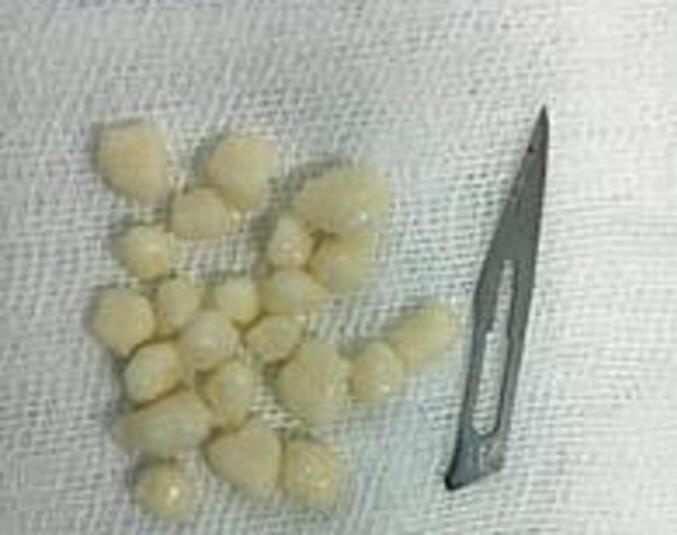
An orthopedist evaluated a 26-year-old man with a history of recurring ankle trauma over a period of 6 months, swelling and pain. An ankle radiograph revealed calcified foreign bodies, while arthrography displayed enlarged oval bodies with contrasting features and intra-articular effusion encapsulated heterogeneously. Subsequent arthroscopy, following contrast injection, revealed intra-articular contrast effusion. During the arthroscopic procedure, multiple osteochondromas were discovered. A synovectomy was performed, and pathological analysis confirmed the presence of synovial chondromatosis.
Synovial chondromatosis of the ankle is an exceptionally rare condition with only a handful of documented cases in the literature. Isolated instances of ankle synovial chondromatosis have been reported, and these cases have been managed using both open and arthroscopic techniques. Arthroscopic intervention offers potential benefits such as improved joint access, reduced morbidity, and quicker rehabilitation and recovery. However, arthroscopic surgery might pose the risk of incomplete synovectomy or residual loose bodies.
Synovial chondromatosis of the ankle represents a chronic and rare ailment. Arthroscopic treatment stands out as an effective and dependable solution, offering various potential advantages over open surgery. This case report is presented due to the rarity and clinical significance of the condition.
20岁的年轻人最易患滑膜软骨瘤病,这是一种罕见病症,其特征为滑膜化生为软骨或骨软骨组织。滑膜软骨瘤病在踝关节极为罕见。
一名骨科医生对一名26岁男性进行了评估,该患者有6个月反复踝关节创伤史,伴有肿胀和疼痛。踝关节X线片显示有钙化异物,关节造影显示椭圆形物体增大,具有不同的特征,关节内积液呈不均匀包裹。随后在注射造影剂后进行关节镜检查,发现关节内有造影剂渗出。在关节镜检查过程中,发现多个骨软骨瘤。进行了滑膜切除术,病理分析证实存在滑膜软骨瘤病。
踝关节滑膜软骨瘤病极为罕见,文献中仅有少数病例记载。已有孤立的踝关节滑膜软骨瘤病病例报告,这些病例采用开放手术和关节镜技术进行治疗。关节镜干预具有潜在优势,如改善关节入路、降低发病率以及更快的康复和恢复。然而,关节镜手术可能存在滑膜切除不完全或残留游离体的风险。
踝关节滑膜软骨瘤病是一种慢性罕见疾病。关节镜治疗是一种有效且可靠的解决方案,与开放手术相比具有多种潜在优势。由于该病症的罕见性和临床意义,特此呈现本病例报告。