Wankhede Sampada, Sahoo Debiprasad, Meshram Aishwarya A, Rane Siddhesh, Borle Nitin
General Surgery, Topiwala National Medical College and Bai Yamunabai Laxman Nair Charitable Hospital, Mumbai, IND.
Gastroenterology, Topiwala National Medical College and Bai Yamunabai Laxman Nair Charitable Hospital, Mumbai, IND.
Cureus. 2024 Oct 2;16(10):e70735. doi: 10.7759/cureus.70735. eCollection 2024 Oct.
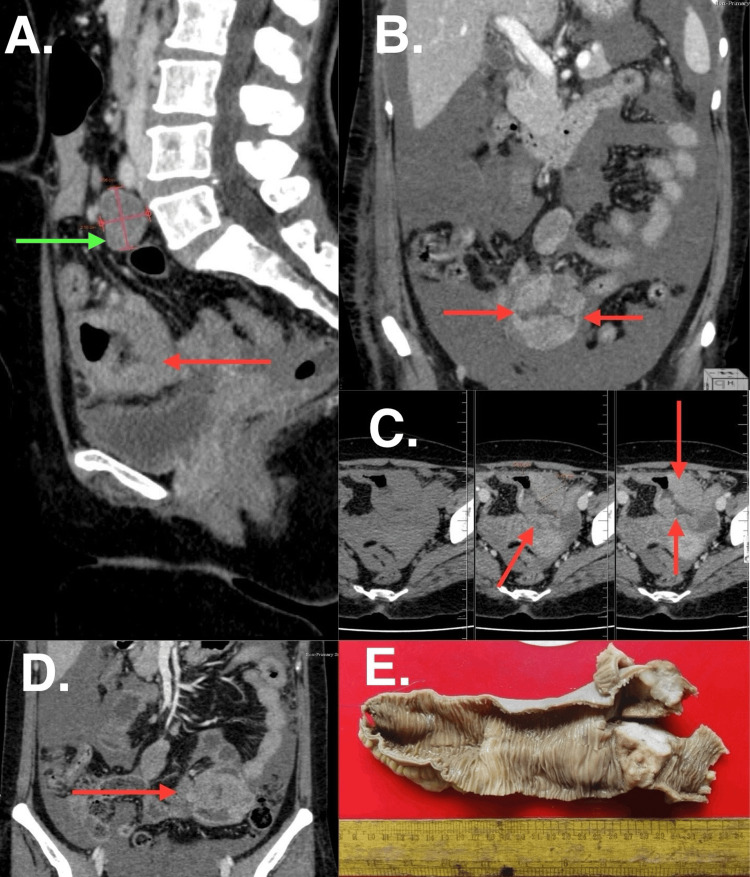
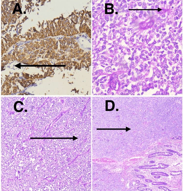
Malignant gastrointestinal neuroectodermal tumor (GNET) are rare malignant mesenchymal tumors. The tumor can present with various symptoms like abdominal pain, anorexia, or small bowel obstruction. Here, we present a case of small intestinal GNET who presented with gastrointestinal bleed and hemoperitoneum, a rare presentation of this disease. This patient was misdiagnosed initially as Crohn's disease and treated for the same. However, non-response to the standard treatment and onset of new symptoms like malena and ascites raised the suspicion of some alternate diagnosis. Exploratory laparotomy showed the presence of hemoperitoneum along with a mass, 100 cm proximal to ileo-cecal junction. She was successfully treated with surgical resection and anastomosis. Histopathology, immunohistochemistry (diffuse positivity for S100 and weak positivity for synaptophysin) and molecular fluorescence in-situ hybridization (FISH) study (translocation involving the chromosomal region 2212.1-q12.2 which harbors gene) confirmed the diagnosis of GNET.
恶性胃肠道神经外胚层肿瘤(GNET)是罕见的恶性间叶组织肿瘤。该肿瘤可表现出各种症状,如腹痛、厌食或小肠梗阻。在此,我们报告一例小肠GNET患者,其表现为胃肠道出血和血腹,这是该疾病的一种罕见表现。该患者最初被误诊为克罗恩病并接受了相应治疗。然而,对标准治疗无反应以及出现黑便和腹水等新症状引发了对其他诊断的怀疑。剖腹探查显示存在血腹以及一个位于回盲部近端100厘米处的肿块。她通过手术切除和吻合术成功治愈。组织病理学、免疫组化(S100弥漫阳性,突触素弱阳性)和分子荧光原位杂交(FISH)研究(涉及22号染色体区域12.1-q12.2的易位,该区域包含 基因)证实了GNET的诊断。