• 文献检索
  • 文档翻译
  • 深度研究
  • 学术资讯
  • Suppr Zotero 插件Zotero 插件
  • 邀请有礼
  • 套餐&价格
  • 历史记录
应用&插件
Suppr Zotero 插件Zotero 插件浏览器插件Mac 客户端Windows 客户端微信小程序
定价
高级版会员购买积分包购买API积分包
服务
文献检索文档翻译深度研究API 文档MCP 服务
关于我们
关于 Suppr公司介绍联系我们用户协议隐私条款
关注我们

Suppr 超能文献

核心技术专利:CN118964589B侵权必究
粤ICP备2023148730 号-1Suppr @ 2026

文献检索

告别复杂PubMed语法,用中文像聊天一样搜索,搜遍4000万医学文献。AI智能推荐,让科研检索更轻松。

立即免费搜索

文件翻译

保留排版,准确专业,支持PDF/Word/PPT等文件格式,支持 12+语言互译。

免费翻译文档

深度研究

AI帮你快速写综述,25分钟生成高质量综述,智能提取关键信息,辅助科研写作。

立即免费体验

我的AIS患者需要哪些影像学检查?提供者偏好的多组调查。

What imaging does my AIS patient need? A multi-group survey of provider preferences.

作者信息

Zheng Jenny L, Li Ying, Hogue Grant, Johnson Megan, Anari Jason B, Regan Maia D, Baldwin Keith D

机构信息

Division of Orthopaedic Surgery, Children's Hospital of Philadelphia, Perelman School of Medicine at the University of Pennsylvania, Philadelphia, PA, USA.

Department of Orthopaedic Surgery, C.S. Mott Children's Hospital, University of Michigan Health, Ann Arbor, MI, USA.

出版信息

Spine Deform. 2025 Mar;13(2):351-359. doi: 10.1007/s43390-024-00995-9. Epub 2024 Nov 4.

DOI:10.1007/s43390-024-00995-9
PMID:39495401
原文链接:https://pmc.ncbi.nlm.nih.gov/articles/PMC11893670/
Abstract

INTRODUCTION

Adolescent idiopathic scoliosis (AIS) is a common diagnosis managed by pediatric orthopedic surgeons with nonoperative radiographic monitoring representing a cornerstone of treatment. Differences in practices and techniques for obtaining radiographic studies contribute to variation, cost of care, and hamper data aggregation. We surveyed several large organizations dedicated to children's orthopedics or scoliosis care to obtain a consensus for radiographic evaluation of AIS.

METHODS

A REDCap-based survey was developed across four institutions and beta-tested by staff and fellows from a single institution. The finalized survey was distributed to members of POSNA, PSSG, and SOSORT, and shared on social media. Participants were asked to rank the importance of various datapoints in radiographic assessment of the spinal deformity, skeletal maturity, and study indications during initial, subsequent, preoperative, and final office visits for AIS. Response rate for the overall group was 26%.

RESULTS

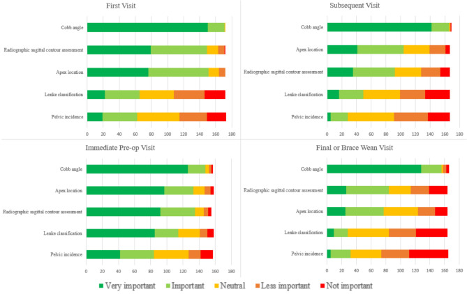
Cobb angle was considered the most important (> 94%) radiographic index across all time points. For positioning, 46% of respondents favored arms bent touching clavicles as the ideal positioning for X-rays, and another 24% favored arms down with palms forward (Table 2). The majority of respondents obtain lateral X-rays at the first visit (99%) and at the preoperative visit (70%). At the preoperative visit, sagittal contour (86%), apex location (85%), and Lenke classification (73%) were considered important factors to record. Flexibility studies are primarily obtained at the preoperative visit (89%) and 81% of respondents prefer bending films as the flexibility technique of choice. Regarding measures of skeletal maturity, Sanders bone age was considered to be the most important by over 70% of respondents across initial, subsequent, preoperative and brace wean visits (Fig. 2). MRIs were obtained routinely by 34% of respondents and only when the patient had a concerning symptom or finding for 67% of respondents.

CONCLUSIONS

Despite large variations in radiographic examination of AIS, large areas of agreement were found. It is important to establish standards for positioning patients, evaluating skeletal maturity, and obtaining assessments including lateral views, flexibility studies, and advanced imaging. Establishing common practices for radiographic evaluation of AIS will allow for less variation in care and for critical questions to be answered through registry formation and large multicenter data collection.

SIGNIFICANCE

This study establishes current practitioner opinion on the radiographic evaluation of the AIS patient. Minimum data sets are useful for data aggregation and answering research questions in the face of data variability.

LEVEL OF EVIDENCE

Level V.

https://cdn.ncbi.nlm.nih.gov/pmc/blobs/4ad7/11893670/da8ddf68837f/43390_2024_995_Fig2_HTML.jpg
https://cdn.ncbi.nlm.nih.gov/pmc/blobs/4ad7/11893670/398921b7bb84/43390_2024_995_Fig1_HTML.jpg
https://cdn.ncbi.nlm.nih.gov/pmc/blobs/4ad7/11893670/da8ddf68837f/43390_2024_995_Fig2_HTML.jpg
https://cdn.ncbi.nlm.nih.gov/pmc/blobs/4ad7/11893670/398921b7bb84/43390_2024_995_Fig1_HTML.jpg
https://cdn.ncbi.nlm.nih.gov/pmc/blobs/4ad7/11893670/da8ddf68837f/43390_2024_995_Fig2_HTML.jpg
摘要

引言

青少年特发性脊柱侧凸(AIS)是儿科骨科医生常见的诊断疾病,非手术影像学监测是治疗的基石。获取影像学研究的方法和技术差异导致了差异、护理成本,并阻碍了数据汇总。我们调查了几个致力于儿童骨科或脊柱侧凸护理的大型组织,以就AIS的影像学评估达成共识。

方法

在四个机构开展了一项基于REDCap的调查,并由一个机构的工作人员和研究员进行了预测试。最终确定的调查分发给了POSNA、PSSG和SOSORT的成员,并在社交媒体上分享。参与者被要求对脊柱畸形、骨骼成熟度的影像学评估以及AIS初次、后续、术前和最终门诊就诊时的研究指征中各种数据点的重要性进行排名。整个群体的回复率为26%。

结果

在所有时间点,Cobb角被认为是最重要的(>94%)影像学指标。对于体位摆放,46%的受访者倾向于双臂弯曲触碰锁骨作为X线检查的理想体位,另有24%的受访者倾向于双臂下垂手掌向前(表2)。大多数受访者在初次就诊(99%)和术前就诊(70%)时获取侧位X线片。在术前就诊时,矢状面轮廓(86%)、顶点位置(85%)和Lenke分型(73%)被认为是重要的记录因素。柔韧性研究主要在术前就诊时进行(89%),81%的受访者更喜欢弯曲位片作为柔韧性检查的首选技术。关于骨骼成熟度的测量,超过70%的受访者在初次、后续、术前和支具撤除就诊时认为Sanders骨龄是最重要的(图2)。34%的受访者常规进行MRI检查,67%的受访者仅在患者有可疑症状或发现时进行检查。

结论

尽管AIS的影像学检查存在很大差异,但仍发现了广泛的共识领域。为患者体位摆放、评估骨骼成熟度以及获取包括侧位片、柔韧性研究和高级影像学检查在内的评估建立标准非常重要。建立AIS影像学评估的通用方法将减少护理差异,并通过建立登记系统和进行大型多中心数据收集来回答关键问题。

意义

本研究确立了当前从业者对AIS患者影像学评估的观点。面对数据变异性,最小数据集对于数据汇总和回答研究问题很有用。

证据水平

V级。

相似文献

1
What imaging does my AIS patient need? A multi-group survey of provider preferences.我的AIS患者需要哪些影像学检查?提供者偏好的多组调查。
Spine Deform. 2025 Mar;13(2):351-359. doi: 10.1007/s43390-024-00995-9. Epub 2024 Nov 4.
2
Adolescent Idiopathic Scoliosis Minimum Data Set: Towards Standardization of Data Elements in History and Physical Examination.青少年特发性脊柱侧凸最小数据集:迈向病史和体格检查数据元素的标准化
Cureus. 2024 Apr 15;16(4):e58332. doi: 10.7759/cureus.58332. eCollection 2024 Apr.
3
A Predictive Model of Progression for Adolescent Idiopathic Scoliosis Based on 3D Spine Parameters at First Visit.基于初次就诊时的三维脊柱参数建立青少年特发性脊柱侧凸进展的预测模型。
Spine (Phila Pa 1976). 2020 May 1;45(9):605-611. doi: 10.1097/BRS.0000000000003316.
4
Reproducibility of sagittal radiographic parameters in adolescent idiopathic scoliosis-a guide to reference values using serial imaging.青少年特发性脊柱侧凸矢状面影像学参数的可重复性——使用系列成像确定参考值的指南
Spine J. 2017 Jun;17(6):830-836. doi: 10.1016/j.spinee.2017.01.001. Epub 2017 Jan 5.
5
The effectiveness of selective thoracic fusion for treating adolescent idiopathic scoliosis: a systematic review protocol.选择性胸椎融合术治疗青少年特发性脊柱侧凸的有效性:一项系统评价方案
JBI Database System Rev Implement Rep. 2015 Nov;13(11):4-16. doi: 10.11124/jbisrir-2015-2338.
6
Role of Clavicle Chest Cage Angle Difference in Predicting Postoperative Shoulder Balance in Lenke 5C Adolescent Idiopathic Scoliosis Patients after Selective Posterior Fusion.锁骨胸廓角差异在预测Lenke 5C型青少年特发性脊柱侧凸患者选择性后路融合术后肩部平衡中的作用
Orthop Surg. 2017 Feb;9(1):86-90. doi: 10.1111/os.12305. Epub 2017 Mar 9.
7
Does the Use of Sanders Staging and Distal Radius and Ulna Classification Avoid Mismatches in Growth Assessment with Risser Staging Alone?单用 Risser 分期评估时,使用 Sanders 分期和桡骨远端及尺骨分类是否可避免生长评估失配?
Clin Orthop Relat Res. 2021 Nov 1;479(11):2516-2530. doi: 10.1097/CORR.0000000000001817.
8
Immediate postoperative coronal imbalance in Lenke 5 and Lenke 6 adolescent idiopathic scoliosis: Is it predictable?后路矫形手术即刻冠状面失平衡在 Lenke 5 和 Lenke 6 型青少年特发性脊柱侧凸中的表现:可预测吗?
Eur Spine J. 2019 Sep;28(9):2042-2052. doi: 10.1007/s00586-019-06019-9. Epub 2019 May 30.
9
Sagittal balance in adolescent idiopathic scoliosis: radiographic study of spino-pelvic compensation after surgery.青少年特发性脊柱侧凸的矢状面平衡:术后脊柱-骨盆代偿的影像学研究
Eur Spine J. 2013 Nov;22 Suppl 6(Suppl 6):S859-67. doi: 10.1007/s00586-013-3018-8. Epub 2013 Sep 24.
10
When Should We Wean Bracing for Adolescent Idiopathic Scoliosis?青少年特发性脊柱侧凸支具治疗何时应停止?
Clin Orthop Relat Res. 2019 Sep;477(9):2145-2157. doi: 10.1097/CORR.0000000000000781.

本文引用的文献

1
Do hypokyphotic adolescent idiopathic scoliosis patients treated with Ponte osteotomy obtain a better clinical efficacy? A preliminary retrospective study.后路楔形截骨术治疗青少年特发性脊柱侧凸患者的临床疗效如何?一项初步的回顾性研究。
J Orthop Surg Res. 2022 Nov 16;17(1):491. doi: 10.1186/s13018-022-03390-0.
2
Benchmarking surgical indications for adolescent idiopathic scoliosis across time, region, and patient population: a study of 4229 cases.跨时间、跨地区和跨患者人群比较青少年特发性脊柱侧凸的手术适应证:一项 4229 例病例的研究。
Spine Deform. 2022 Jul;10(4):833-840. doi: 10.1007/s43390-022-00480-1. Epub 2022 Mar 8.
3
Specific sagittal alignment patterns are already present in mild adolescent idiopathic scoliosis.
轻度青少年特发性脊柱侧凸已经存在特定的矢状位排列模式。
Eur Spine J. 2021 Jul;30(7):1881-1887. doi: 10.1007/s00586-021-06772-w. Epub 2021 Feb 27.
4
Validation of the National Institute of Neurological Disorders and Stroke Spinal Cord Injury MRI Common Data Elements Instrument.验证国家神经疾病与中风研究所脊髓损伤 MRI 通用数据元素工具。
AJNR Am J Neuroradiol. 2021 Apr;42(4):787-793. doi: 10.3174/ajnr.A7000. Epub 2021 Feb 11.
5
Influence of curve morphology and location on the efficacy of rigid conservative treatment in patients with adolescent idiopathic scoliosis.曲线形态和位置对青少年特发性脊柱侧凸患者刚性保守治疗效果的影响。
Bone Joint J. 2021 Feb;103-B(2):373-381. doi: 10.1302/0301-620X.103B2.BJJ-2020-1113.R2.
6
Scoliosis Screening.脊柱侧凸筛查。
J Am Acad Orthop Surg. 2021 May 1;29(9):370-379. doi: 10.5435/JAAOS-D-20-00356.
7
Sanders stage 7b: Using the appearance of the ulnar physis improves decision-making for brace weaning in patients with adolescent idiopathic scoliosis.桑德斯 7b 期:使用尺骨骨骺的外观可改善青少年特发性脊柱侧凸患者支具脱卸的决策。
Bone Joint J. 2021 Jan;103-B(1):141-147. doi: 10.1302/0301-620X.103B1.BJJ-2020-1240.R1.
8
Assessment of the plane of maximum curvature for patients with adolescent idiopathic scoliosis via computed tomography.通过计算机断层扫描评估青少年特发性脊柱侧凸患者的最大曲率平面。
Prosthet Orthot Int. 2020 Oct;44(5):298-304. doi: 10.1177/0309364620941297. Epub 2020 Jul 22.
9
It is not just about the frontal plane: sagittal parameters impact curve progression in AIS patients undergoing brace treatment.这不仅仅关乎额状面:矢状面参数会影响接受支具治疗的青少年特发性脊柱侧弯(AIS)患者的侧弯进展。
Spine Deform. 2020 Oct;8(5):921-929. doi: 10.1007/s43390-020-00122-4. Epub 2020 Apr 27.
10
High Risk of Mismatch Between Sanders and Risser Staging in Adolescent Idiopathic Scoliosis: Are We Guiding Treatment Using the Wrong Classification?青少年特发性脊柱侧凸中桑德斯(Sanders)和里塞尔(Risser)分期不匹配的高风险:我们是否在用错误的分类指导治疗?
J Pediatr Orthop. 2020 Feb;40(2):60-64. doi: 10.1097/BPO.0000000000001135.