Department of Ophthalmology, Tianjin Medical University General Hospital, Tianjin, China.
School of Medicine, Nankai University, Tianjin, China.
Invest Ophthalmol Vis Sci. 2024 Nov 4;65(13):8. doi: 10.1167/iovs.65.13.8.
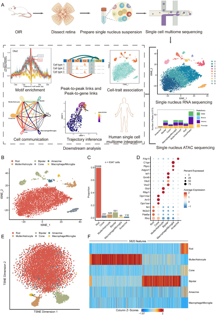
Retinal neovascularization poses heightened risks of vision loss and blindness. Despite its clinical significance, the molecular mechanisms underlying the pathogenesis of retinal neovascularization remain elusive. This study utilized single-cell multiomics profiling in an oxygen-induced retinopathy (OIR) model to comprehensively investigate the intricate molecular landscape of retinal neovascularization.
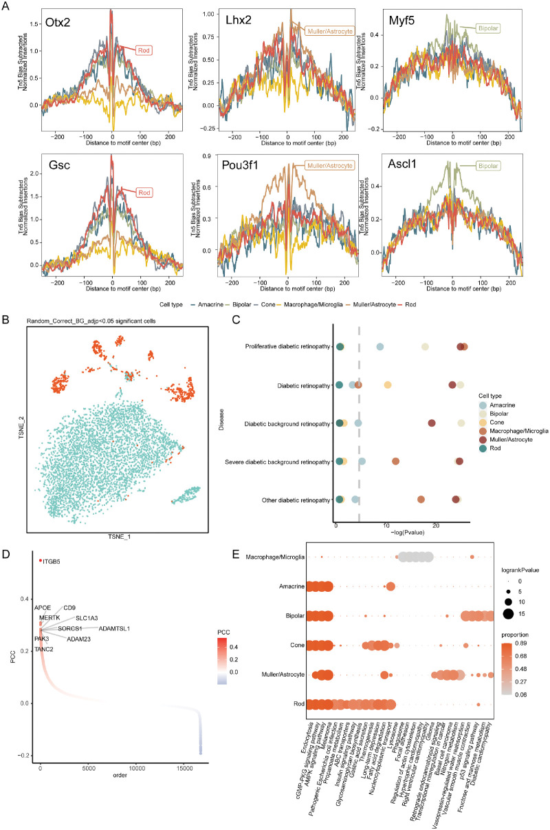
Mice were exposed to hyperoxia to induce the OIR model, and retinas were isolated for nucleus isolation. The cellular landscape of the single-nucleus suspensions was extensively characterized through single-cell multiomics sequencing. Single-cell data were integrated with genome-wide association study (GWAS) data to identify correlations between ocular cell types and diabetic retinopathy. Cell communication analysis among cells was conducted to unravel crucial ligand-receptor signals. Trajectory analysis and dynamic characterization of Müller cells were performed, followed by integration with human retinal data for pathway analysis.
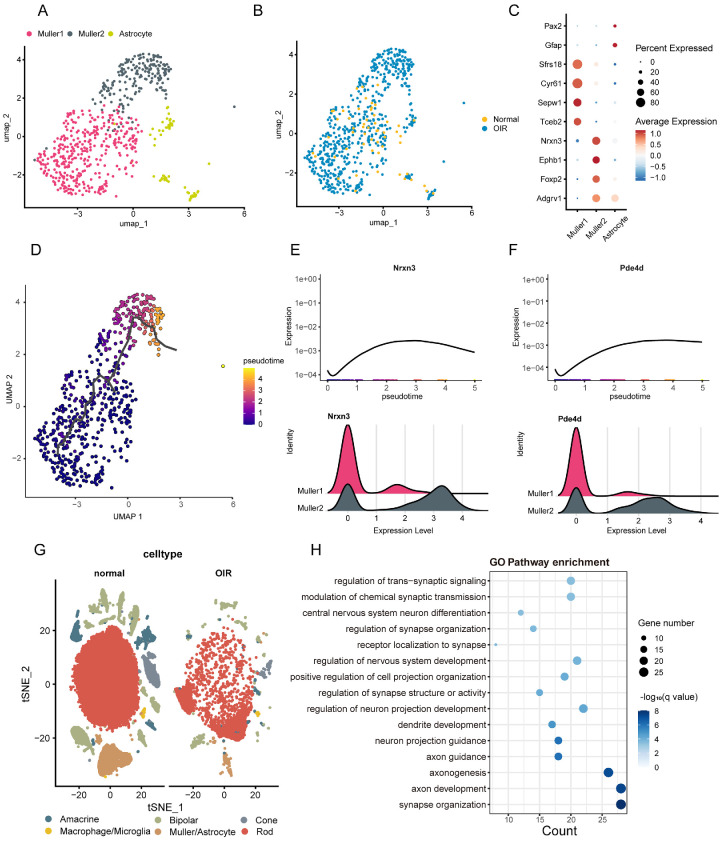
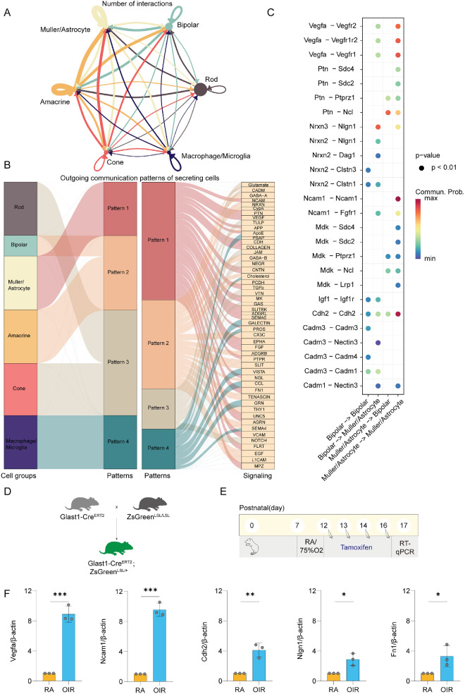
The multiomics dataset revealed six major ocular cell classes, with Müller cells/astrocytes showing significant associations with proliferative diabetic retinopathy (PDR). Cell communication analysis highlighted pathways that are associated with vascular proliferation and neurodevelopment, such as Vegfa-Vegfr2, Igf1-Igf1r, Nrxn3-Nlgn1, and Efna5-Epha4. Trajectory analysis identified a subset of Müller cells expressing genes linked to photoreceptor degeneration. Multiomics data integration further unveiled positively regulated genes in OIR Müller cells/astrocytes associated with axon development and neurotransmitter transmission.
This study significantly advances our understanding of the intricate cellular and molecular mechanisms underlying retinal neovascularization, emphasizing the pivotal role of Müller cells. The identified pathways provide valuable insights into potential therapeutic targets for PDR, offering promising directions for further research and clinical interventions.
视网膜新生血管会增加视力丧失和失明的风险。尽管其具有临床意义,但视网膜新生血管发病机制的分子机制仍难以捉摸。本研究利用单细胞多组学分析在氧诱导的视网膜病变(OIR)模型中,全面研究视网膜新生血管的复杂分子景观。
将小鼠暴露于高氧环境中以诱导 OIR 模型,并分离视网膜进行核分离。通过单细胞多组学测序广泛表征单细胞悬浮液的细胞景观。将单细胞数据与全基因组关联研究(GWAS)数据集成,以确定眼部细胞类型与糖尿病视网膜病变之间的相关性。对细胞间的细胞通讯进行分析,以揭示关键的配体-受体信号。对 Müller 细胞进行轨迹分析和动态特征分析,并与人类视网膜数据进行整合以进行途径分析。
多组学数据集揭示了六种主要的眼部细胞类型,其中 Müller 细胞/星形胶质细胞与增殖性糖尿病视网膜病变(PDR)具有显著相关性。细胞通讯分析突出了与血管增殖和神经发育相关的途径,如 Vegfa-Vegfr2、Igf1-Igf1r、Nrxn3-Nlgn1 和 Efna5-Epha4。轨迹分析确定了一组表达与光感受器退行性变相关基因的 Müller 细胞。多组学数据集成进一步揭示了 OIR Müller 细胞/星形胶质细胞中与轴突发育和神经递质传递相关的正向调节基因。
本研究显著提高了我们对视网膜新生血管发病机制中复杂细胞和分子机制的理解,强调了 Müller 细胞的关键作用。确定的途径为 PDR 的潜在治疗靶点提供了有价值的见解,为进一步的研究和临床干预提供了有希望的方向。