• 文献检索
  • 文档翻译
  • 深度研究
  • 学术资讯
  • Suppr Zotero 插件Zotero 插件
  • 邀请有礼
  • 套餐&价格
  • 历史记录
应用&插件
Suppr Zotero 插件Zotero 插件浏览器插件Mac 客户端Windows 客户端微信小程序
定价
高级版会员购买积分包购买API积分包
服务
文献检索文档翻译深度研究API 文档MCP 服务
关于我们
关于 Suppr公司介绍联系我们用户协议隐私条款
关注我们

Suppr 超能文献

核心技术专利:CN118964589B侵权必究
粤ICP备2023148730 号-1Suppr @ 2026

文献检索

告别复杂PubMed语法,用中文像聊天一样搜索,搜遍4000万医学文献。AI智能推荐,让科研检索更轻松。

立即免费搜索

文件翻译

保留排版,准确专业,支持PDF/Word/PPT等文件格式,支持 12+语言互译。

免费翻译文档

深度研究

AI帮你快速写综述,25分钟生成高质量综述,智能提取关键信息,辅助科研写作。

立即免费体验

乳腺影像报告和数据系统(BI-RADS)乳腺增强磁共振成像(CEM)词典的阅片者间一致性

Inter-reader agreement of the BI-RADS CEM lexicon.

作者信息

Zarcaro Calogero, Santonocito Ambra, Zeitouni Layla, Ferrara Francesca, Kapetas Panagiotis, Milos Ruxandra-Iulia, Helbich Thomas H, Baltzer Pascal A T, Clauser Paola

机构信息

Department of Biomedicine, Neuroscience and Advanced Diagnostic (Bi.N.D.), University Hospital "Policlinico P. Giaccone", Palermo, Italy.

Department of Biomedical Imaging and Image-Guided Therapy, Medical University of Vienna, Vienna, Austria.

出版信息

Eur Radiol. 2025 May;35(5):2378-2386. doi: 10.1007/s00330-024-11176-7. Epub 2024 Nov 6.

DOI:10.1007/s00330-024-11176-7
PMID:39505735
原文链接:https://pmc.ncbi.nlm.nih.gov/articles/PMC12021736/
Abstract

PURPOSE

The purpose of this study was to assess the inter-reader agreement of the breast imaging reporting and data system (BI-RADS) contrast-enhanced mammography (CEM) lexicon.

MATERIALS AND METHODS

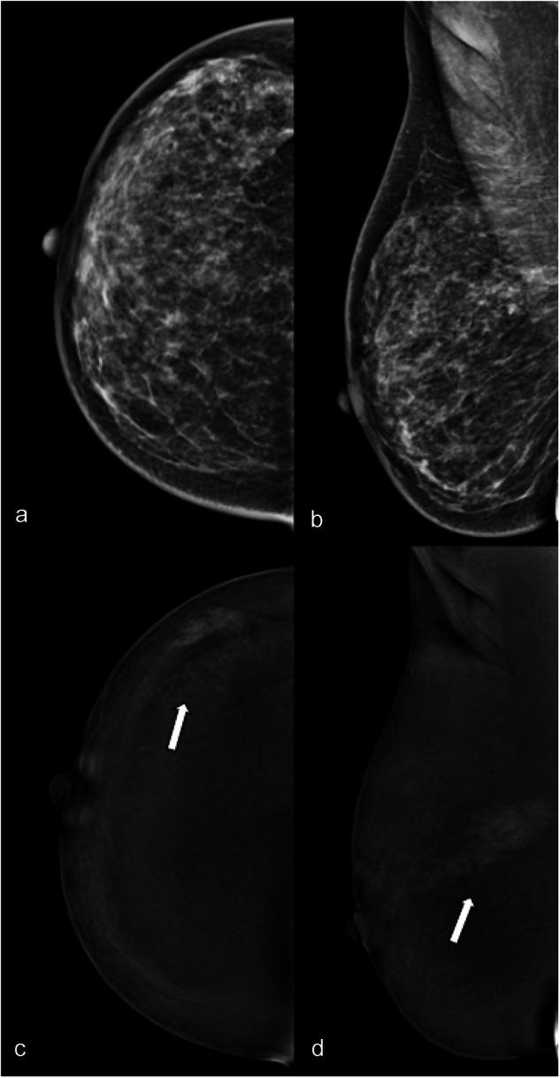
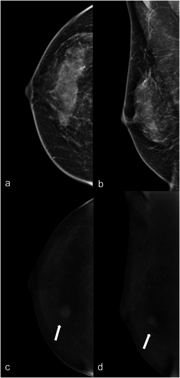
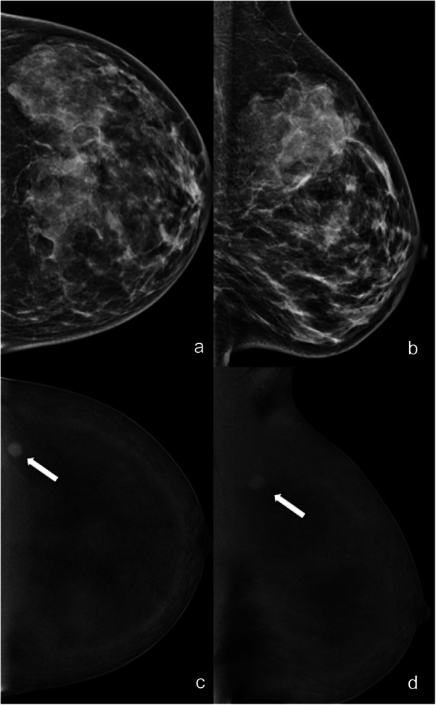
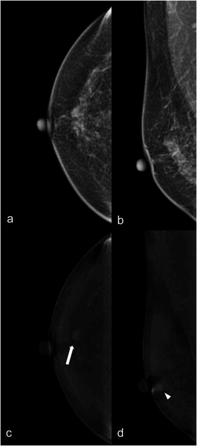
In this IRB-approved, single-center, retrospective study, three breast radiologists, each with different levels of experience, reviewed 462 lesions in 421 routine clinical CEM according to the fifth edition of the BI-RADS lexicon for mammography and to the first version of the BI-RADS lexicon for CEM. Readers were blinded to patient outcomes and evaluated breast and lesion features on low-energy (LE) images (breast density, type of lesion, associated architectural distortion), lesion features on recombined (RC) images (type of enhancement, characteristic of mass enhancement, non-mass enhancement or enhancing asymmetry), and provided a final BI-RADS assessment. The inter-reader agreement was calculated for each evaluated feature using Fleiss' kappa coefficient. Sensitivity and specificity were calculated.

RESULTS

The inter-reader agreement was moderate to substantial for breast density (ĸ = 0.569), type of lesion on LE images (ĸ = 0.654), and type of enhancement (ĸ = 0.664). There was a moderate to substantial agreement on CEM mass enhancement descriptors. The agreement was fair to moderate for non-mass enhancement and enhancing asymmetry descriptors. Inter-reader agreement for LE and LE with RC BI-RADS assessment was moderate (ĸ = 0.421) and fair (ĸ = 0.364). Diagnostic performance was good and comparable for all readers.

CONCLUSION

Inter-reader agreement of the CEM lexicon was moderate to substantial for most features. There was a low agreement for some RC descriptors, such as non-mass enhancement and enhancing asymmetry, and BI-RADS assessment, but this did not impact the diagnostic performance.

KEY POINTS

Question Data on the reproducibility and inter-reader agreement for the first version of the BI-RADS lexicon dedicated to CEM are missing. Finding The inter-reader agreement for the lexicon was overall substantial to moderate, but it was lower for the descriptors for non-mass enhancement and enhancing asymmetry. Clinical relevance A common lexicon simplifies communication between specialists in clinical practice. The good inter-reader agreement confirms the effectiveness of the CEM-BIRADS in ensuring consistent communication. Detailed definitions of some descriptors (non-mass, enhancing asymmetry) are needed to ensure higher agreements.

摘要

目的

本研究旨在评估乳腺影像报告和数据系统(BI-RADS)对比增强乳腺X线摄影(CEM)词汇表在不同阅片者之间的一致性。

材料与方法

在这项经机构审查委员会(IRB)批准的单中心回顾性研究中,三名经验水平不同的乳腺放射科医生,根据乳腺X线摄影BI-RADS词汇表第五版和CEM的BI-RADS词汇表第一版,对421例常规临床CEM中的462个病灶进行了评估。阅片者对患者的预后情况不知情,并在低能量(LE)图像上评估乳腺和病灶特征(乳腺密度、病灶类型、相关的结构扭曲),在重组(RC)图像上评估病灶特征(强化类型、肿块强化特征、非肿块强化或强化不对称),并给出最终的BI-RADS评估。使用Fleiss卡方系数计算每个评估特征在不同阅片者之间的一致性。计算敏感性和特异性。

结果

对于乳腺密度(κ = 0.569)、LE图像上的病灶类型(κ = 0.654)和强化类型(κ = 0.664),不同阅片者之间的一致性为中等至高度。对于CEM肿块强化描述符存在中等至高度的一致性。对于非肿块强化和强化不对称描述符,一致性为一般至中等。LE以及LE与RC的BI-RADS评估在不同阅片者之间的一致性为中等(κ = 0.421)和一般(κ = 0.364)。所有阅片者的诊断性能良好且相当。

结论

CEM词汇表在大多数特征上,不同阅片者之间的一致性为中等至高度。对于一些RC描述符,如非肿块强化和强化不对称以及BI-RADS评估,一致性较低,但这并未影响诊断性能。

关键点

问题 缺少关于专门用于CEM的BI-RADS词汇表第一版再现性和不同阅片者之间一致性的数据。发现 词汇表在不同阅片者之间的一致性总体上为高度至中等,但非肿块强化和强化不对称描述符的一致性较低。临床意义 通用词汇表简化了临床实践中专家之间的沟通。良好的不同阅片者之间的一致性证实了CEM-BIRADS在确保一致沟通方面的有效性。需要对一些描述符(非肿块、强化不对称)进行详细定义以确保更高的一致性。

https://cdn.ncbi.nlm.nih.gov/pmc/blobs/815b/12021736/cc85a4c1f353/330_2024_11176_Fig4_HTML.jpg
https://cdn.ncbi.nlm.nih.gov/pmc/blobs/815b/12021736/3b3ed16a5ef6/330_2024_11176_Fig1_HTML.jpg
https://cdn.ncbi.nlm.nih.gov/pmc/blobs/815b/12021736/b9249684719e/330_2024_11176_Fig2_HTML.jpg
https://cdn.ncbi.nlm.nih.gov/pmc/blobs/815b/12021736/1552f424414d/330_2024_11176_Fig3_HTML.jpg
https://cdn.ncbi.nlm.nih.gov/pmc/blobs/815b/12021736/cc85a4c1f353/330_2024_11176_Fig4_HTML.jpg

相似文献

1
Inter-reader agreement of the BI-RADS CEM lexicon.乳腺影像报告和数据系统(BI-RADS)乳腺增强磁共振成像(CEM)词典的阅片者间一致性
Eur Radiol. 2025 May;35(5):2378-2386. doi: 10.1007/s00330-024-11176-7. Epub 2024 Nov 6.
2
A head-to-head comparison of breast lesion's conspicuity at contrast-enhanced mammography and contrast-enhanced MRI.对比增强乳腺钼靶摄影和对比增强磁共振成像对乳腺病变的显示能力的直接比较。
Eur Radiol. 2025 Jun;35(6):3070-3079. doi: 10.1007/s00330-024-11195-4. Epub 2024 Dec 3.
3
Assessment of Pectoralis muscle invasion using combined DBT and contrast-enhanced mammography: Retrospective multi-reader study.使用数字化乳腺断层合成(DBT)与对比增强乳腺X线摄影相结合评估胸大肌侵犯:回顾性多阅片者研究
Curr Probl Diagn Radiol. 2025 Apr 17. doi: 10.1067/j.cpradiol.2025.04.005.
4
Inter-reader reliability of MRI and CT Node Reporting and Data System 1.0 (Node-RADS) for mesorectal lymph nodes in rectal cancer.磁共振成像(MRI)和计算机断层扫描(CT)的直肠系膜淋巴结报告及数据系统1.0(Node-RADS)在直肠癌直肠系膜淋巴结评估中的阅片者间可靠性。
Acta Radiol. 2025 Jul;66(7):687-694. doi: 10.1177/02841851251322887. Epub 2025 Jul 15.
5
Screening for Breast Cancer with Contrast-enhanced Mammography as an Alternative to MRI: SCEMAM Trial Results.对比增强乳腺钼靶筛查作为MRI替代方法用于乳腺癌筛查:SCEMAM试验结果
Radiology. 2025 Jun;315(3):e242634. doi: 10.1148/radiol.242634.
6
Validation of the American College of Radiology Bone Reporting and Data System™ (ACR Bone-RADS™) for classifying osteolytic bone tumors.美国放射学会骨报告与数据系统™(ACR骨影像报告和数据系统™,ACR Bone-RADS™)对溶骨性骨肿瘤进行分类的验证。
Skeletal Radiol. 2025 Feb 3. doi: 10.1007/s00256-025-04881-x.
7
Analysis of ILD-RADS reproducibility and validity: Can different degrees of experience affect the interpretation?间质性肺疾病影像报告和数据系统(ILD-RADS)的可重复性和有效性分析:不同程度的经验会影响解读结果吗?
Eur J Radiol. 2025 Mar;184:111961. doi: 10.1016/j.ejrad.2025.111961. Epub 2025 Jan 30.
8
Bone Reporting and Data System on MRI (Bone-RADS-MRI): a validation study by four readers on 275 cases from three local and two public databases.MRI骨报告与数据系统(骨影像报告和数据系统 - MRI):由四位阅片者对来自三个本地数据库和两个公共数据库的275例病例进行的验证研究
Insights Imaging. 2025 Jul 17;16(1):155. doi: 10.1186/s13244-025-02040-3.
9
Variation within and between digital pathology and light microscopy for the diagnosis of histopathology slides: blinded crossover comparison study.数字病理学与光学显微镜检查在组织病理学切片诊断中的内部及相互间差异:双盲交叉对比研究
Health Technol Assess. 2025 Jul;29(30):1-75. doi: 10.3310/SPLK4325.
10
Inter-reader reliability of Ovarian-Adnexal Reporting and Data System US: a systematic review and meta-analysis.卵巢附件报告和数据系统超声检查的阅片者间可靠性:一项系统评价和荟萃分析
Abdom Radiol (NY). 2025 Jan 22. doi: 10.1007/s00261-025-04813-2.

引用本文的文献

1
An Updated Systematic Review and Meta-Analysis of Diagnostic Accuracy of Dynamic Contrast Enhancement and Diffusion-Weighted MRI in Differentiating Benign and Malignant Non-Mass Enhancement Lesions.动态对比增强和扩散加权磁共振成像鉴别良性和恶性非肿块强化病变诊断准确性的最新系统评价和荟萃分析
J Clin Med. 2025 Jun 30;14(13):4628. doi: 10.3390/jcm14134628.
2
18F-FDG PET/CT Semiquantitative and Radiomic Features for Assessing Pathologic Axillary Lymph Node Status in Clinical Stage I-III Breast Cancer Patients: A Systematic Review.18F-FDG PET/CT半定量和影像组学特征用于评估临床I-III期乳腺癌患者腋窝病理淋巴结状态:一项系统综述
Curr Oncol. 2025 May 23;32(6):300. doi: 10.3390/curroncol32060300.

本文引用的文献

1
Addition of Contrast-enhanced Mammography to Tomosynthesis for Breast Cancer Detection in Women with a Personal History of Breast Cancer: Prospective TOCEM Trial Interim Analysis.增强型乳腺 X 光摄影术联合断层合成乳腺摄影术在有乳腺癌个人病史的女性乳腺癌检测中的应用:前瞻性 TOCEM 试验中期分析。
Radiology. 2024 Apr;311(1):e231991. doi: 10.1148/radiol.231991.
2
Contrast-enhanced Mammography versus Contrast-enhanced Breast MRI: A Systematic Review and Meta-Analysis.对比增强乳腺 X 线摄影与对比增强乳腺 MRI:系统评价和荟萃分析。
Radiology. 2022 Oct;305(1):94-103. doi: 10.1148/radiol.212530. Epub 2022 Jun 7.
3
A Score to Predict the Malignancy of a Breast Lesion Based on Different Contrast Enhancement Patterns in Contrast-Enhanced Spectral Mammography.
基于对比增强光谱乳腺摄影中不同对比增强模式预测乳腺病变恶性程度的评分系统
Cancers (Basel). 2022 Sep 5;14(17):4337. doi: 10.3390/cancers14174337.
4
Contrast-enhanced Mammography: A Systematic Review and Meta-Analysis of Diagnostic Performance.对比增强乳腺摄影术:诊断性能的系统评价和荟萃分析。
Radiology. 2022 Mar;302(3):568-581. doi: 10.1148/radiol.211412. Epub 2021 Dec 14.
5
Contrast-enhanced Mammography: State of the Art.对比增强乳腺摄影:现状。
Radiology. 2021 Apr;299(1):36-48. doi: 10.1148/radiol.2021201948. Epub 2021 Mar 2.
6
Diagnostic Performance of MRI, Molecular Breast Imaging, and Contrast-enhanced Mammography in Women with Newly Diagnosed Breast Cancer.MRI、分子乳腺成像和对比增强乳腺摄影在新诊断乳腺癌女性中的诊断性能。
Radiology. 2019 Dec;293(3):531-540. doi: 10.1148/radiol.2019190887. Epub 2019 Oct 29.
7
Comparison of inter- and intra-observer variability of breast density assessments using the fourth and fifth editions of Breast Imaging Reporting and Data System.使用《乳腺影像报告和数据系统》第四版和第五版对乳腺密度评估中观察者间和观察者内变异性的比较。
Eur J Radiol Open. 2018 Apr 20;5:67-72. doi: 10.1016/j.ejro.2018.04.002. eCollection 2018.
8
Contrast Enhanced Spectral Mammography: A Review.对比增强光谱乳腺摄影术:综述
Semin Ultrasound CT MR. 2018 Feb;39(1):70-79. doi: 10.1053/j.sult.2017.08.005. Epub 2017 Aug 24.
9
Application of BI-RADS Descriptors in Contrast-Enhanced Dual-Energy Mammography: Comparison with MRI.BI-RADS描述符在双能对比增强乳腺摄影中的应用:与MRI的比较。
Breast Care (Basel). 2017 Sep;12(4):212-216. doi: 10.1159/000478899. Epub 2017 Aug 17.
10
Revisiting Nonmass Enhancement in Breast MRI: Analysis of Outcomes and Follow-Up Using the Updated BI-RADS Atlas.重新审视乳腺MRI中的非肿块强化:使用更新版BI-RADS图谱分析结果及随访情况。
AJR Am J Roentgenol. 2017 Nov;209(5):1178-1184. doi: 10.2214/AJR.17.18086. Epub 2017 Aug 23.