Schmidt Tuany R, Mármora Belkiss C, Brochado Fernanda T, Gonçalves Lucas, Campos Paloma S, Lamers Marcelo L, Araújo Aurigena A de, Medeiros Caroline A C X de, Ribeiro Susana B, Martins Marco A T, Pilar Emily F S, Martins Manoela D, Wagner Vivian P
Department of Pathology, Faculty of Dentistry, Universidade Federal do Rio Grande do Sul, Porto Alegre, RS, Brazil.
Department of Pathology, Faculty of Dentistry, Universidade Federal do Rio Grande do Sul, Porto Alegre, RS, Brazil; Department of Pediatric Dentistry, Orthodontics and Public Health, Faculty of Dentistry, Universidade de São Paulo, Bauru, SP, Brazil.
An Bras Dermatol. 2025 Jan-Feb;100(1):54-62. doi: 10.1016/j.abd.2024.02.008. Epub 2024 Nov 8.
The clinical advantages of light-emitting diode (LED) therapy in skin healing and its underlying mechanism remain subjects of ongoing debate.
This study aims to explore the impact of LED therapy on normal skin keratinocytes (HaCaT) and in the repair of full-thickness dorsal wounds in Wistar rats.
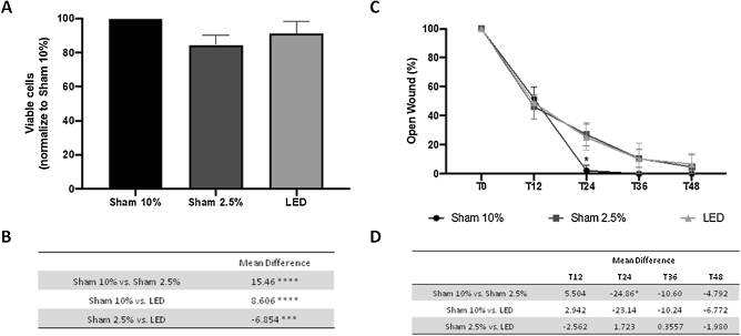
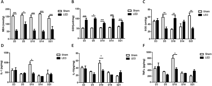
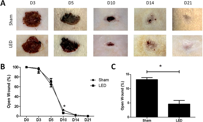
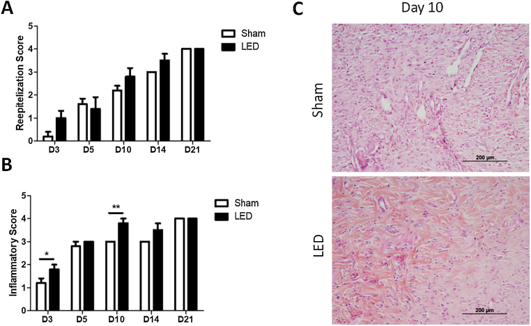
HaCaT cell viability (SRB assay) and migration (scratch assay) were assessed under LED therapy, comparing stress conditions (2.5% FBS) with sham irradiation and optimal conditions (10% FBS). In vivo, 50 rats with induced wounds were divided into Sham and LED (daily treatment) groups. Euthanasia occurred at 3, 5, 10, 14, and 21 days for clinical, morphological, oxidative stress (MDA, SOD, and GSH), and cytokine analyses (IL-1β, IL-10, TNF-α).
LED therapy significantly enhanced keratinocytes viability compared to sham irradiation, with minimal impact on cell migration. Clinical benefits were prominent on day 10, influencing inflammation progression and resolution on days 3 and 10. Re-epithelization remained unaffected. Reduced MDA and increased GSH levels were observed throughout, while SOD levels varied temporally. Notably, on day 10, LED significantly decreased IL-1β, IL-10, and TNF-α.
Although translational, clinical trial confirmation of observed benefits is warranted.
LED therapy expedites cutaneous healing in the experimental model, primarily modulating inflammation and enhancing antioxidant activity.
发光二极管(LED)疗法在皮肤愈合方面的临床优势及其潜在机制仍是持续争论的话题。
本研究旨在探讨LED疗法对正常皮肤角质形成细胞(HaCaT)的影响以及对Wistar大鼠背部全层伤口修复的作用。
在LED疗法下,通过磺酰罗丹明B(SRB)法评估HaCaT细胞活力,划痕试验评估细胞迁移能力,将应激条件(2.5%胎牛血清)与假照射及最佳条件(10%胎牛血清)进行比较。在体内,将50只诱导伤口的大鼠分为假手术组和LED(每日治疗)组。在第3、5、10、14和21天实施安乐死,进行临床、形态学、氧化应激(丙二醛、超氧化物歧化酶和谷胱甘肽)及细胞因子分析(白细胞介素-1β、白细胞介素-10、肿瘤坏死因子-α)。
与假照射相比,LED疗法显著提高了角质形成细胞活力,对细胞迁移影响最小。在第10天临床益处显著,对第3天和第10天的炎症进展和消退有影响。再上皮化未受影响。整个过程中丙二醛水平降低,谷胱甘肽水平升高,而超氧化物歧化酶水平随时间变化。值得注意的是,在第