Lee Christine U, Hesley Gina K, Pierson Taylor A, Higgins Rebecca L, Urban Matthew W
Department of Radiology, Division of Breast Imaging and Intervention, Mayo Clinic, Rochester, MN, USA.
Department of Radiology, Division of Radiology Research, Mayo Clinic, Rochester, MN, USA.
Transl Breast Cancer Res. 2024 Oct 29;5:28. doi: 10.21037/tbcr-24-30. eCollection 2024.
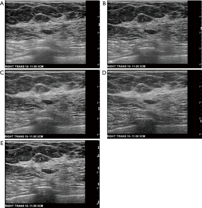
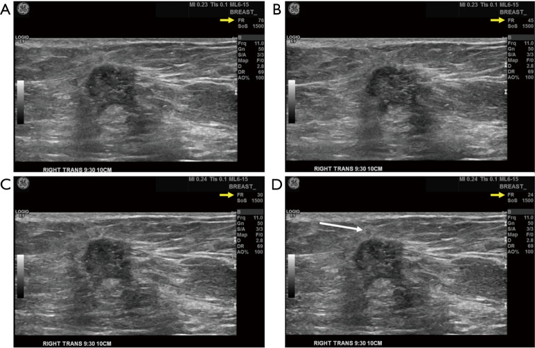
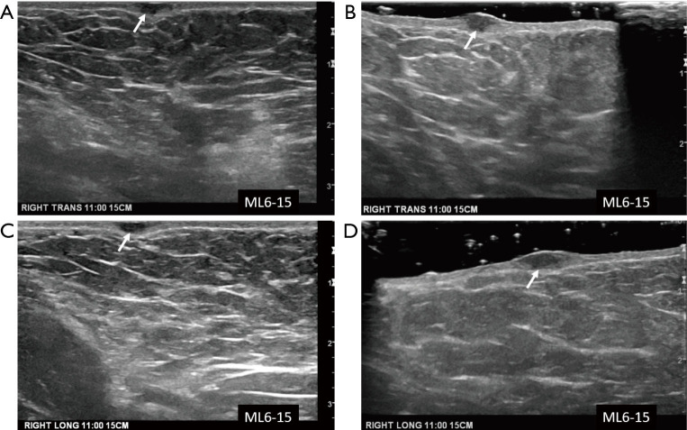
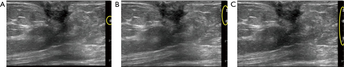
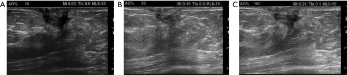
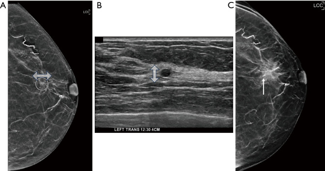
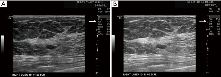
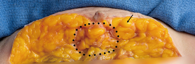
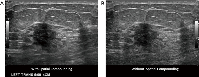
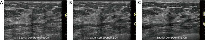
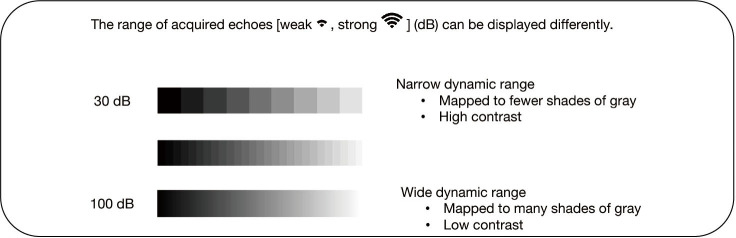
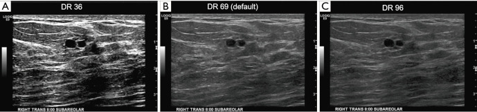
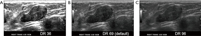
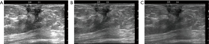
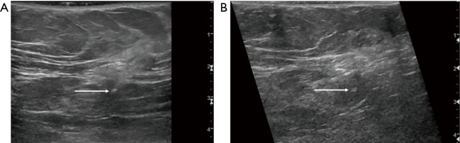
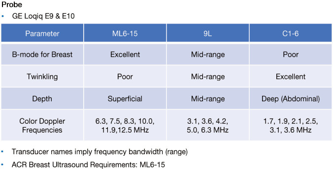
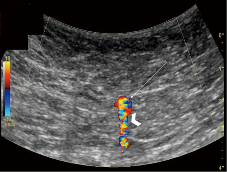
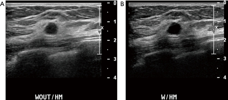
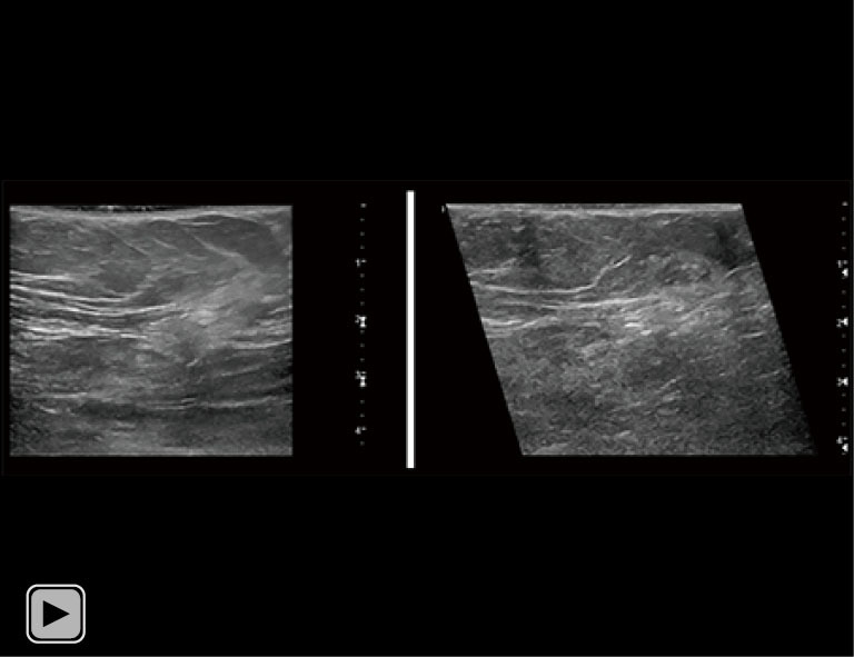
Breast ultrasound utilizes various scanning techniques to acquire optimal images for diagnostic evaluation. During interventional procedures, such as ultrasound-guided biopsies or preoperative localizations, knowledgeable and purposeful scanning adjustments are critical for successfully identifying the targeted mass or biopsy marker or clip. While most ultrasound scanning parameters are similar across different vendors, detailed descriptions specifically addressing the scanning parameters-often referred to as "knobology"- for breast ultrasound is at best limited in the literature. This review highlights ten key operator-controlled adjustments (including transducer selection, beam focusing, power, depth, gain and time gain compensation, harmonic imaging, spatial compounding, dynamic range, beam steering, and color Doppler) that significantly influence image quality in breast ultrasound. Each adjustment is accompanied by an "In practice" section providing examples and practical tips on implementation. The last topic discusses color Doppler which is generally used in breast ultrasound for evaluating the vascularity of a finding. Color Doppler, or more specifically, color Doppler twinkling, can be leveraged as a technique to detect certain breast biopsy markers that are challenging to detect by conventional B-mode ultrasound. While the cause of color Doppler twinkling is still under active investigation, twinkling is a clinically well-known, compelling ultrasound feature typically described with kidney stones. A step-by-step guide on how to use color Doppler twinkling to detect these markers is provided.
乳腺超声利用多种扫描技术获取最佳图像用于诊断评估。在介入操作过程中,如超声引导下活检或术前定位,知识丰富且有针对性的扫描调整对于成功识别目标肿块、活检标记物或夹子至关重要。虽然不同厂家的大多数超声扫描参数相似,但文献中针对乳腺超声扫描参数(通常称为“旋钮操作”)的详细描述非常有限。本综述重点介绍了十个关键的操作员控制调整(包括换能器选择、波束聚焦、功率、深度、增益和时间增益补偿、谐波成像、空间复合、动态范围、波束控制和彩色多普勒),这些调整会显著影响乳腺超声的图像质量。每个调整都附有一个“实际操作”部分,提供实施示例和实用技巧。最后一个主题讨论了彩色多普勒,它通常用于乳腺超声中评估发现病变的血管情况。彩色多普勒,或者更具体地说,彩色多普勒闪烁,可以作为一种技术来检测某些传统B超难以检测到的乳腺活检标记物。虽然彩色多普勒闪烁的原因仍在积极研究中,但闪烁是一种临床上众所周知且引人注目的超声特征,通常用于描述肾结石。本文提供了一份关于如何使用彩色多普勒闪烁来检测这些标记物的分步指南。