Zhou Zheng, Cheng Xu, Yang Fan, Zhang Zhihua, Liu Kaiping, Zhang Xin, Huang Hongjie, Wang Jianquan
Department of Sports Medicine, Institute of Sports Medicine of Peking University, Peking University Third Hospital, Beijing, China.
Key Laboratory of Astronaut Health Center, China Astronaut Research and Training Center, Beijing, China.
Ann Jt. 2024 Oct 30;9:37. doi: 10.21037/aoj-24-6. eCollection 2024.
Long-term exposure to weightlessness can result in bone and muscle degradation, significantly impacting musculoskeletal function. Recent studies have also indicated damage to articular cartilage due to weightlessness. This study aims to observe the effects of simulated weightlessness on the cartilage microstructure of the quadriceps muscle and the muscular knee joint in rats.
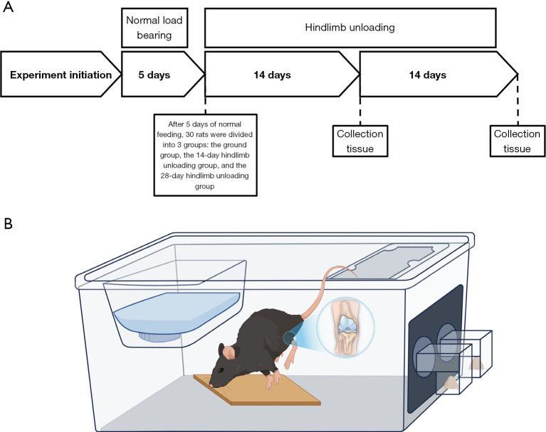
A total of 30 rats were used in this study, of which 20 rats were subjected to simulated weightlessness by tail suspension, which may be suitable for clinical long-term bedridden patients. At 14 and 28 days, the microscopic morphology of knee cartilage and quadriceps femoris muscle was observed by transmission electron microscopy, and the collagen and water content of cartilage was evaluated by magnetic resonance imaging. The mitochondrial activity of knee muscle and the levels of inflammatory factors in synovial fluid were detected by enzyme-linked immunosorbent assay (ELISA). Biomechanical and histological evaluation of cartilage was performed.
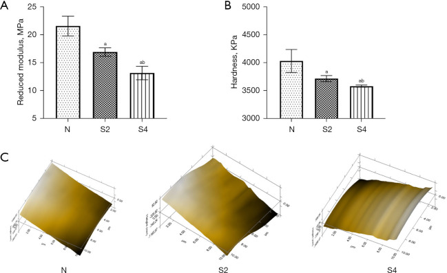
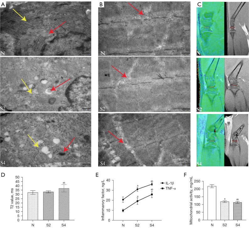
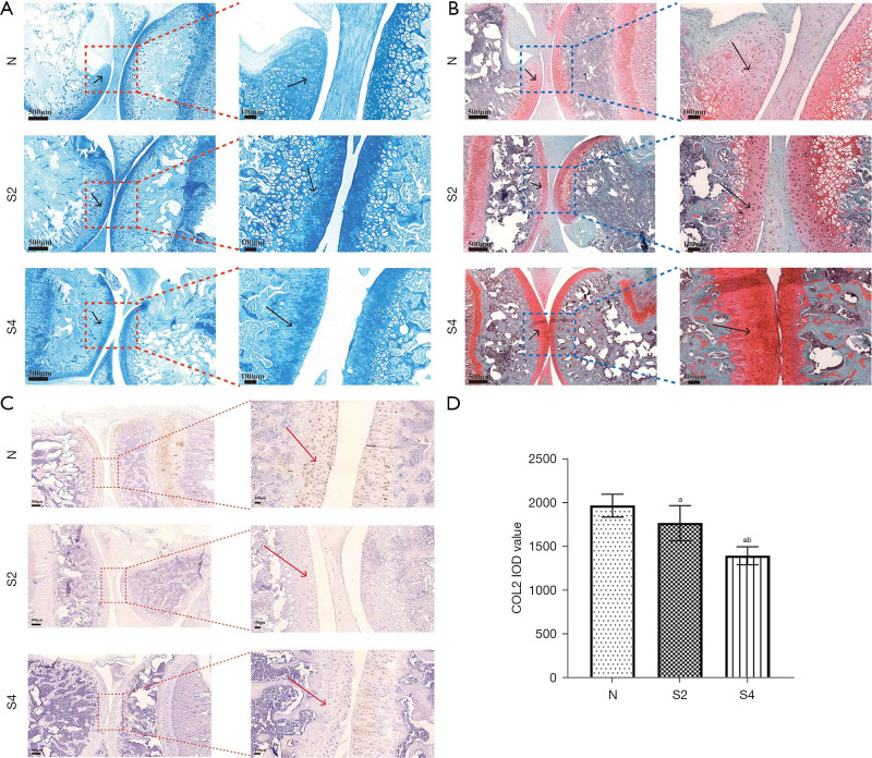
On day 14, T2 mapping revealed no significant loading effect. However, transmission electron microscopy revealed altered mitochondrial inner membrane structure in cartilage, with vacuolization, disrupted endoplasmic reticulum, alongside mitochondrial ultrastructural damage in muscle. ELISA results showed that a large number of mitochondria in muscle were inactivated, and the levels of inflammatory factors in synovial fluid were increased. The staining results showed slight fracture of the cartilage surface and the type II collagen-positive cells were reduced. Nanoindentation showed that the cartilage microsurface was uneven, and the elastic modulus and hardness were decreased. On day 28, T2 mapping analysis indicated increased cartilage T2 values. Transmission electron microscopy showed alterations in the structure of the mitochondrial inner membrane in cartilage, severe vacuolization, disrupted endoplasmic reticulum, and substantial mitochondrial damage in muscle tissue. Muscle mitochondrial activity was markedly decreased, inflammatory factors levels were elevated, and the cartilage surface exhibited severe damage. The type II collagen positive cells were further reduced, the micro-surface of cartilage was uneven, and the elastic modulus and hardness were significantly decreased.
The weightless environment resulted in the damage of endoplasmic reticulum and mitochondria of cartilage, mitochondrial damage of quadriceps muscle, inactivation of muscle mitochondria (P=0.01), increased intra-articular inflammation (P=0.01), decreased elastic modulus and hardness (P=0.03), and damaged cartilage surface, which aggravated cartilage degeneration.
长期暴露于失重环境会导致骨骼和肌肉退化,对肌肉骨骼功能产生重大影响。最近的研究还表明失重会对关节软骨造成损害。本研究旨在观察模拟失重对大鼠股四头肌和膝关节软骨微观结构的影响。
本研究共使用30只大鼠,其中20只大鼠通过尾部悬吊进行模拟失重处理,这可能适用于临床长期卧床患者。在第14天和第28天,通过透射电子显微镜观察膝关节软骨和股四头肌的微观形态,并用磁共振成像评估软骨的胶原蛋白和水分含量。通过酶联免疫吸附测定(ELISA)检测膝关节肌肉的线粒体活性和滑液中炎症因子的水平。对软骨进行生物力学和组织学评估。
在第14天,T2映射显示无明显负荷效应。然而,透射电子显微镜显示软骨中线粒体内膜结构改变,出现空泡化、内质网破坏,同时肌肉中线粒体超微结构受损。ELISA结果显示肌肉中大量线粒体失活,滑液中炎症因子水平升高。染色结果显示软骨表面有轻微骨折,II型胶原阳性细胞减少。纳米压痕显示软骨微观表面不均匀,弹性模量和硬度降低。在第28天,T2映射分析表明软骨T2值增加。透射电子显微镜显示软骨中线粒体内膜结构改变明显,空泡化严重,内质网破坏,肌肉组织中线粒体大量损伤。肌肉线粒体活性明显降低,炎症因子水平升高,软骨表面出现严重损伤。II型胶原阳性细胞进一步减少,软骨微观表面不均匀,弹性模量和硬度显著降低。
失重环境导致软骨内质网和线粒体损伤、股四头肌线粒体损伤、肌肉线粒体失活(P=0.01)、关节内炎症增加(P=0.01)、弹性模量和硬度降低(P=0.03)以及软骨表面受损,加重了软骨退变。