Department of Biology, Indiana University-Purdue University Indianapolis, Indianapolis, IN, 46202, USA.
Stark Neurosciences Research Institute, Indiana University School of Medicine, Indianapolis, IN, 46202, USA.
Fluids Barriers CNS. 2024 Nov 14;21(1):90. doi: 10.1186/s12987-024-00593-x.
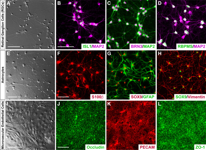
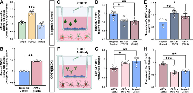
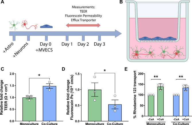
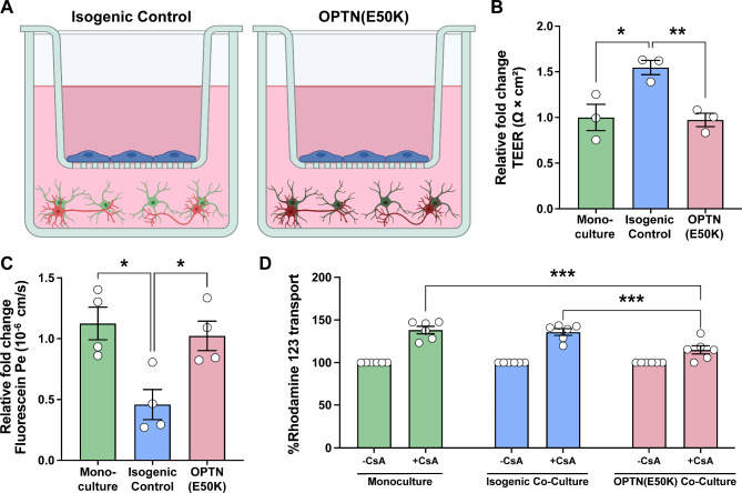
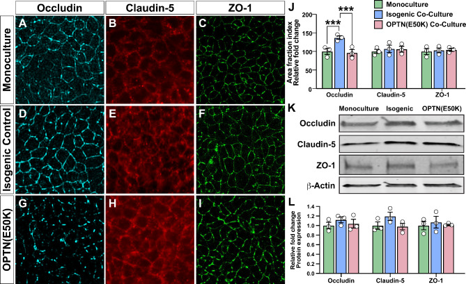
Glaucoma is a neurodegenerative disease that results in the degeneration of retinal ganglion cells (RGCs) and subsequent loss of vision. While RGCs are the primary cell type affected in glaucoma, neighboring cell types selectively modulate RGCs to maintain overall homeostasis. Among these neighboring cell types, astrocytes, microvascular endothelial cells (MVECs), and pericytes coordinate with neurons to form the neurovascular unit that provides a physical barrier to limit the passage of toxic materials from the blood into neural tissue. Previous studies have demonstrated that these barrier properties may be compromised in the progression of glaucoma, yet mechanisms by which this happens have remained incompletely understood. Thus, the goals of this study were to adapt a human pluripotent stem cell (hPSC)-based model of the neurovascular unit to the study of barrier integrity relevant to glaucoma. To achieve this, hPSCs were differentiated into the cell types that contribute to this barrier, including RGCs, astrocytes, and MVECs, then assembled into an established Transwell-insert model. The ability of these cell types to contribute to an in vitro barrier model was tested for their ability to recapitulate characteristic barrier properties. Results revealed that barrier properties of MVECs were enhanced when cultured in the presence of RGCs and astrocytes compared to MVECs cultured alone. Conversely, the versatility of this system to model aspects of barrier dysfunction relevant to glaucoma was tested using an hPSC line with a glaucoma-specific Optineurin (E50K) mutation as well as a paired isogenic control, where MVECs then exhibited reduced barrier integrity. To identify factors that could result in barrier dysfunction, results revealed an increased expression of TGFβ2 in glaucoma-associated OPTN(E50K) astrocytes, indicating a potential role for TGFβ2 in disease manifestation. To test this hypothesis, we explored the ability to modulate exogenous TGFβ2 in both isogenic control and OPTN(E50K) experimental conditions. Collectively, the results of this study indicated that the repurposing of this in vitro barrier model for glaucoma reliably mimicked some aspects of barrier dysfunction, and may serve as a platform for drug discovery, as well as a powerful in vitro model to test the consequences of barrier dysfunction upon RGCs in glaucoma.
青光眼是一种神经退行性疾病,导致视网膜神经节细胞(RGCs)的变性和随后的视力丧失。虽然 RGCs 是青光眼主要受影响的细胞类型,但邻近的细胞类型选择性地调节 RGCs 以维持整体内稳态。在这些邻近的细胞类型中,星形胶质细胞、微血管内皮细胞(MVECs)和周细胞与神经元协调形成神经血管单元,为限制有毒物质从血液进入神经组织提供物理屏障。先前的研究表明,这些屏障特性在青光眼的进展中可能受损,但发生这种情况的机制仍不完全清楚。因此,本研究的目标是适应基于人多能干细胞(hPSC)的神经血管单元模型,以研究与青光眼相关的屏障完整性。为了实现这一目标,将 hPSC 分化为有助于该屏障的细胞类型,包括 RGCs、星形胶质细胞和 MVECs,然后组装到已建立的 Transwell 插入模型中。这些细胞类型在体外屏障模型中的能力通过它们重现特征性屏障特性的能力进行了测试。结果表明,与单独培养的 MVECs 相比,在存在 RGCs 和星形胶质细胞的情况下培养的 MVECs 的屏障特性得到增强。相反,该系统用于模拟与青光眼相关的屏障功能障碍的各个方面的多功能性通过使用具有青光眼特异性 Optineurin(E50K)突变的 hPSC 系以及配对的同基因对照进行了测试,其中 MVECs 表现出降低的屏障完整性。为了确定可能导致屏障功能障碍的因素,结果表明在与青光眼相关的 OPTN(E50K)星形胶质细胞中 TGFβ2 的表达增加,表明 TGFβ2 在疾病表现中可能发挥作用。为了验证这一假设,我们在同基因对照和 OPTN(E50K)实验条件下探索了调节外源性 TGFβ2 的能力。总的来说,这项研究的结果表明,该体外屏障模型的重新用于青光眼可靠地模拟了一些屏障功能障碍的方面,并且可以作为药物发现的平台,以及用于测试青光眼对 RGCs 产生的屏障功能障碍后果的强大体外模型。