Department of Pharmacology and Neuroscience, North Texas Eye Research Institute, University of North Texas Health Science Center, Fort Worth, TX 76107, USA.
Int J Mol Sci. 2022 Jun 21;23(13):6883. doi: 10.3390/ijms23136883.
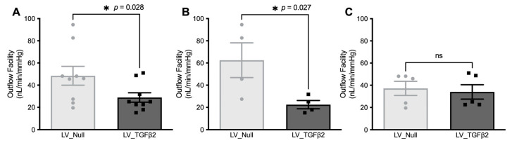
Glaucoma is a multifactorial disease leading to irreversible blindness. Primary open-angle glaucoma (POAG) is the most common form and is associated with the elevation of intraocular pressure (IOP). Reduced aqueous humor (AH) outflow due to trabecular meshwork (TM) dysfunction is responsible for IOP elevation in POAG. Extracellular matrix (ECM) accumulation, actin cytoskeletal reorganization, and stiffening of the TM are associated with increased outflow resistance. Transforming growth factor (TGF) β2, a profibrotic cytokine, is known to play an important role in the development of ocular hypertension (OHT) in POAG. An appropriate mouse model is critical in understanding the underlying molecular mechanism of TGFβ2-induced OHT. To achieve this, TM can be targeted with recombinant viral vectors to express a gene of interest. Lentiviruses (LV) are known for their tropism towards TM with stable transgene expression and low immunogenicity. We, therefore, developed a novel mouse model of IOP elevation using LV gene transfer of active human TGFβ2 in the TM. We developed an LV vector-encoding active hTGFβ2 under the control of a cytomegalovirus (CMV) promoter. Adult C57BL/6J mice were injected intravitreally with LV expressing null or hTGFβ2. We observed a significant increase in IOP 3 weeks post-injection compared to control eyes with an average delta change of 3.3 mmHg. IOP stayed elevated up to 7 weeks post-injection, which correlated with a significant drop in the AH outflow facility (40.36%). Increased expression of active TGFβ2 was observed in both AH and anterior segment samples of injected mice. The morphological assessment of the mouse TM region via hematoxylin and eosin (H&E) staining and direct ophthalmoscopy examination revealed no visible signs of inflammation or other ocular abnormalities in the injected eyes. Furthermore, transduction of primary human TM cells with LV_hTGFβ2 exhibited alterations in actin cytoskeleton structures, including the formation of F-actin stress fibers and crossed-linked actin networks (CLANs), which are signature arrangements of actin cytoskeleton observed in the stiffer fibrotic-like TM. Our study demonstrated a mouse model of sustained IOP elevation via lentiviral gene delivery of active hTGFβ2 that induces TM dysfunction and outflow resistance.
青光眼是一种多因素疾病,可导致不可逆转的失明。原发性开角型青光眼(POAG)是最常见的形式,与眼内压(IOP)升高有关。小梁网(TM)功能障碍导致房水(AH)流出减少,是 POAG 中 IOP 升高的原因。细胞外基质(ECM)积累、肌动蛋白细胞骨架重排和 TM 变硬与流出阻力增加有关。转化生长因子(TGF)β2 是一种促纤维化细胞因子,已知在 POAG 中眼高压(OHT)的发展中起重要作用。适当的小鼠模型对于理解 TGFβ2 诱导的 OHT 的潜在分子机制至关重要。为此,可以使用重组病毒载体靶向 TM 来表达感兴趣的基因。慢病毒(LV)以其对 TM 的趋向性、稳定的转基因表达和低免疫原性而闻名。因此,我们使用 LV 基因转导 TM 中活性人 TGFβ2 开发了一种新的 IOP 升高的小鼠模型。我们开发了一种 LV 载体,在巨细胞病毒(CMV)启动子的控制下表达活性 hTGFβ2。成年 C57BL/6J 小鼠经玻璃体腔内注射表达无效或 hTGFβ2 的 LV。与对照眼相比,注射后 3 周时 IOP 显著升高,平均差值为 3.3mmHg。IOP 升高持续到注射后 7 周,与 AH 流出率(40.36%)显著下降相关。在注射小鼠的 AH 和眼前段样本中观察到活性 TGFβ2 的表达增加。通过苏木精和伊红(H&E)染色和直接检眼镜检查对小鼠 TM 区域的形态评估显示,注射眼无炎症或其他眼部异常的可见迹象。此外,LV_hTGFβ2 转导原代人 TM 细胞后,肌动蛋白细胞骨架结构发生改变,包括 F-肌动蛋白应力纤维和交联肌动蛋白网络(CLANs)的形成,这是在更硬的纤维化样 TM 中观察到的肌动蛋白细胞骨架的特征排列。我们的研究表明,通过慢病毒基因传递活性 hTGFβ2 可诱导 TM 功能障碍和流出阻力增加,从而建立持续升高的 IOP 小鼠模型。