Department of Obstetrics and Gynecology, Peking University People's Hospital, No. 11 Xizhimen South Str., Xicheng District, Beijing, 100044, China.
J Ovarian Res. 2024 Nov 14;17(1):225. doi: 10.1186/s13048-024-01547-5.
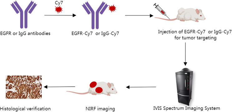
Near-infrared fluorescence (NIRF) imaging is an excellent choice for image-guided surgery due to its simple operation and non-invasiveness. Developing tumor-specific fluorescent molecular probes is key to fluorescence imaging-guided surgery. EGFR (epidermal growth factor receptor) is closely related to the proliferation and growth of tumor cells and is highly expressed in epithelial ovarian cancer (EOC). The study aims to construct a NIR fluorescent molecular probe using cetuximab (an EGFR monoclonal antibody) and investigate its feasibility for targeting EOC in vivo through fluorescence imaging.
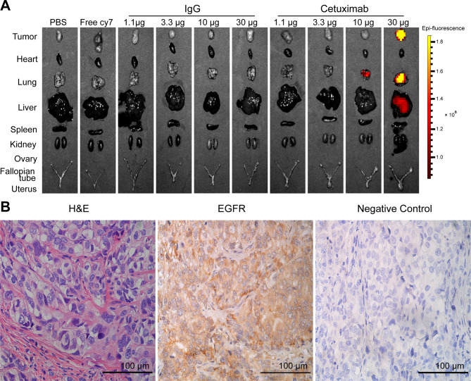
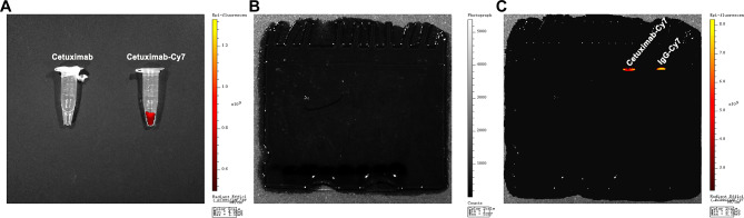
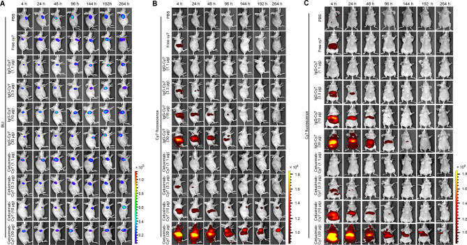
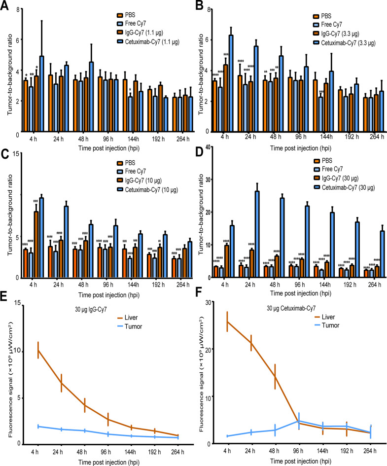
We determined the expression of EGFR in EOC. NIR fluorescent molecular probe with cetuximab (cetuximab-Cy7) was chemically engineered and identified. The subcutaneous xenografted tumor model of EOC was induced using SKOV3-Luc cell line with positive expression of EGFR. Cetuximab-Cy7 was used for in vivo fluorescence imaging, and phosphate-buffered saline, free Cy7 dye and mouse isotype immunoglobulin G-Cy7 were used as controls. NIRF imaging system was performed to study the distribution and targeting of the probes. Tumors were imaged in situ and ex vivo, and fluorescent intensity was quantified. Resected specimens were analyzed to confirm diagnosis, and immunohistochemical (IHC) staining was used to identify EGFR expression.
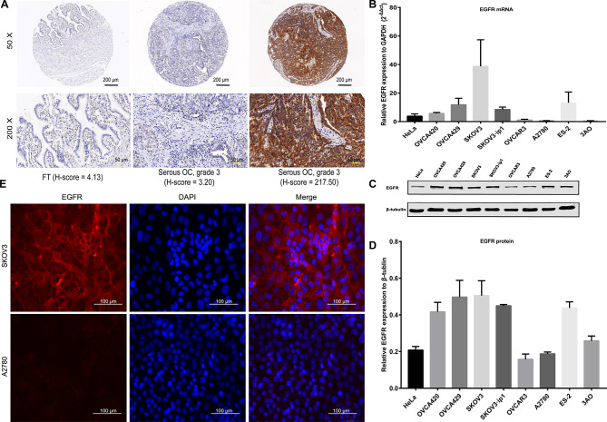
EGFR expression was increased in EOC tissues than fallopian tube tissues. The high expression of EGFR was significantly correlated with well-differentiation, residual lesions ≤ 1 cm, no recurrence and increased survival. NIRF imaging showed that the cetuximab-Cy7 enabled detection of tumor lesions in EOC-bearing mice with the optimal dose of 30 µg. The suitable imaging time window may be 24-96 h post-injection. Ex vivo fluorescence imaging indicated that fluorescent signal was mainly detected in the tumor and the lung. IHC results confirmed that xenografts were EGFR positive.
Cetuximab-Cy7 can specifically target the tumors of EOC xenografted nude mice. This research lays the foundation for future studies on EOC surgery navigation.
近红外荧光(NIRF)成像是一种出色的选择,因其操作简单、非侵入性而广泛应用于影像引导手术。开发肿瘤特异性荧光分子探针是荧光成像引导手术的关键。表皮生长因子受体(EGFR)与肿瘤细胞的增殖和生长密切相关,在卵巢上皮癌(EOC)中高度表达。本研究旨在构建一种使用西妥昔单抗(一种 EGFR 单克隆抗体)的 NIR 荧光分子探针,并通过荧光成像研究其在体内靶向 EOC 的可行性。
我们确定了 EOC 中 EGFR 的表达。通过化学工程方法构建并鉴定了携带西妥昔单抗的 NIR 荧光分子探针(西妥昔单抗-Cy7)。使用 EGFR 阳性表达的 SKOV3-Luc 细胞系诱导卵巢上皮癌皮下移植瘤模型。使用西妥昔单抗-Cy7 进行体内荧光成像,磷酸盐缓冲盐水、游离 Cy7 染料和鼠同型免疫球蛋白 G-Cy7 作为对照。使用 NIRF 成像系统研究探针的分布和靶向性。对原位和离体肿瘤进行成像,并定量荧光强度。对切除标本进行分析以确认诊断,并进行免疫组化(IHC)染色以鉴定 EGFR 表达。
与输卵管组织相比,EOC 组织中 EGFR 表达增加。EGFR 的高表达与分化程度高、残留病灶≤1cm、无复发和生存率提高显著相关。NIRF 成像显示,西妥昔单抗-Cy7 可在携带 EOC 的小鼠中检测到肿瘤病变,最佳剂量为 30μg。合适的成像时间窗口可能是注射后 24-96 小时。离体荧光成像表明,荧光信号主要在肿瘤和肺部检测到。IHC 结果证实异种移植物为 EGFR 阳性。
西妥昔单抗-Cy7 可特异性靶向 EOC 裸鼠异种移植瘤。这项研究为未来的 EOC 手术导航研究奠定了基础。