• 文献检索
  • 文档翻译
  • 深度研究
  • 学术资讯
  • Suppr Zotero 插件Zotero 插件
  • 邀请有礼
  • 套餐&价格
  • 历史记录
应用&插件
Suppr Zotero 插件Zotero 插件浏览器插件Mac 客户端Windows 客户端微信小程序
定价
高级版会员购买积分包购买API积分包
服务
文献检索文档翻译深度研究API 文档MCP 服务
关于我们
关于 Suppr公司介绍联系我们用户协议隐私条款
关注我们

Suppr 超能文献

核心技术专利:CN118964589B侵权必究
粤ICP备2023148730 号-1Suppr @ 2026

文献检索

告别复杂PubMed语法,用中文像聊天一样搜索,搜遍4000万医学文献。AI智能推荐,让科研检索更轻松。

立即免费搜索

文件翻译

保留排版,准确专业,支持PDF/Word/PPT等文件格式,支持 12+语言互译。

免费翻译文档

深度研究

AI帮你快速写综述,25分钟生成高质量综述,智能提取关键信息,辅助科研写作。

立即免费体验

甲状腺相关眼病的眼眶计算机断层扫描影像学特征。

Orbital computed tomography imaging characteristics of thyroid-associated ophthalmopathy.

机构信息

The Affiliated Eye Hospital, Jiangxi Medical College, Nanchang University, No. 463 Bayi Avenue, Nanchang City, 330006, Jiangxi Province, China.

Jiangxi Clinical Research Center for Ophthalmic Disease, Nanchang, Jiangxi, China.

出版信息

Sci Rep. 2024 Nov 22;14(1):28960. doi: 10.1038/s41598-024-76624-2.

DOI:10.1038/s41598-024-76624-2
PMID:39578469
原文链接:https://pmc.ncbi.nlm.nih.gov/articles/PMC11584846/
Abstract

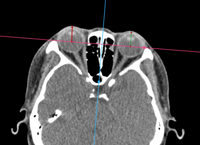
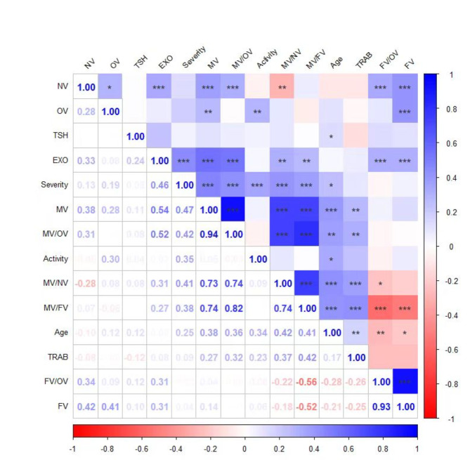
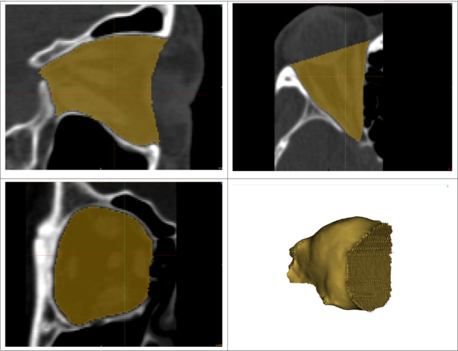
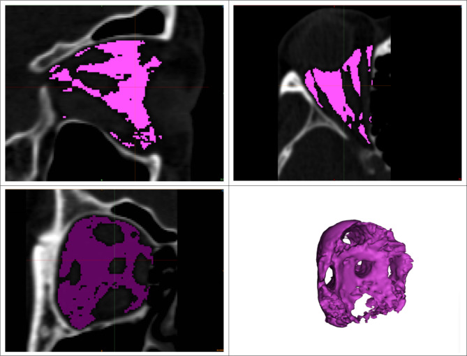
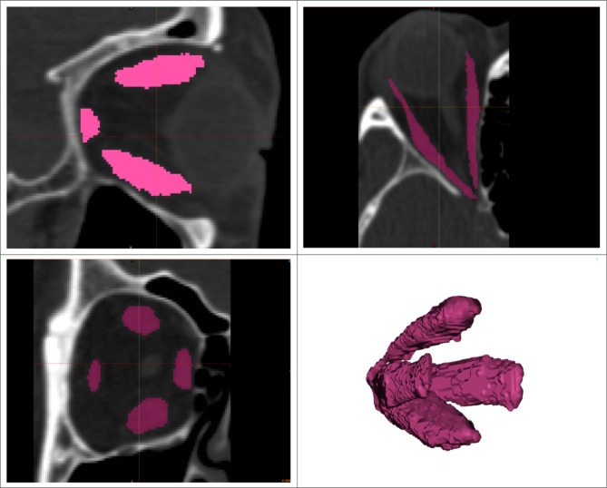
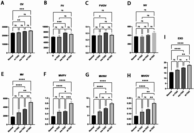
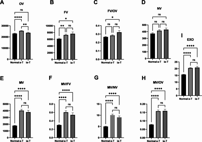
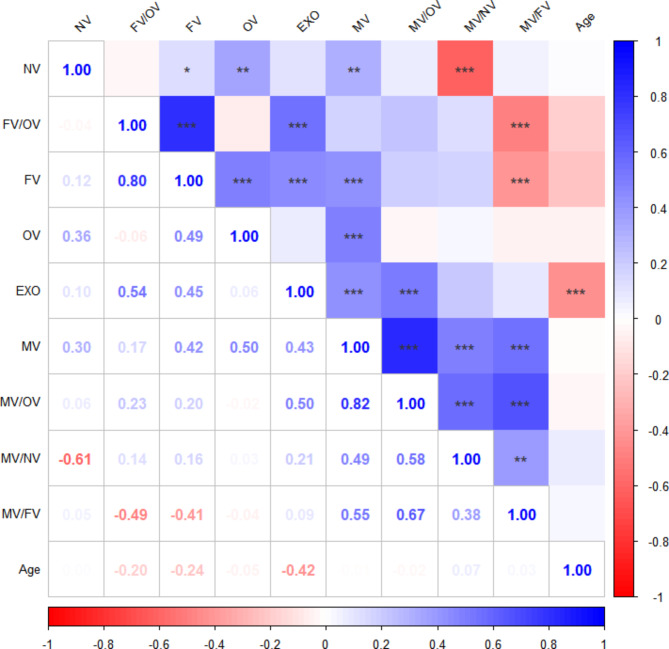
This study aims to investigate the distribution characteristics and correlations of orbital soft tissue volume expansion in patients with thyroid-associated ophthalmopathy (TAO) by analyzing orbital computed tomography (CT) data and to determine the most appropriate parameters and corresponding cut-off values for imaging classification. Patients with TAO who met the inclusion and exclusion criteria, along with those with lacrimal duct obstruction requiring orbital CT examination, were included in the study. Raw CT data were imported into Mimics and RadiAnt software for analysis, and measurements of orbital volume, orbital fat volume, extraocular muscle volume, optic nerve volume, and exophthalmos were obtained. The results demonstrated that the combination of Mimics and other softwares for processing orbital CT scans allows for the quantitative analysis of various soft tissue volumes within the orbit. The MV/FV ratio was found to effectively reflect the relative changes in extraocular muscle volume, serving as a valuable indicator for identifying different subtypes of TAO and providing significant clinical reference value.

摘要

本研究旨在通过分析眼眶 CT 数据,探讨甲状腺相关眼病(TAO)患者眼眶软组织容积扩张的分布特征及相关性,并确定影像学分类的最佳参数及相应截断值。纳入符合纳入排除标准的 TAO 患者及需要眼眶 CT 检查的泪道阻塞患者,将原始 CT 数据导入 Mimics 和 RadiAnt 软件进行分析,测量眼眶容积、眶脂容积、眼外肌容积、视神经容积和眼球突出度。结果表明,Mimics 联合其他软件处理眼眶 CT 扫描可以实现眼眶内各种软组织容积的定量分析。MV/FV 比值能有效反映眼外肌容积的相对变化,是鉴别不同亚型 TAO 的有价值指标,具有重要的临床参考价值。

https://cdn.ncbi.nlm.nih.gov/pmc/blobs/963c/11584846/3fd25967c363/41598_2024_76624_Fig9_HTML.jpg
https://cdn.ncbi.nlm.nih.gov/pmc/blobs/963c/11584846/3b22b2321ff2/41598_2024_76624_Fig1_HTML.jpg
https://cdn.ncbi.nlm.nih.gov/pmc/blobs/963c/11584846/ca8d3e3efa0f/41598_2024_76624_Fig2_HTML.jpg
https://cdn.ncbi.nlm.nih.gov/pmc/blobs/963c/11584846/1c6e5e82c23a/41598_2024_76624_Fig3_HTML.jpg
https://cdn.ncbi.nlm.nih.gov/pmc/blobs/963c/11584846/23cab00b1bad/41598_2024_76624_Fig4_HTML.jpg
https://cdn.ncbi.nlm.nih.gov/pmc/blobs/963c/11584846/29275ac9d542/41598_2024_76624_Fig5_HTML.jpg
https://cdn.ncbi.nlm.nih.gov/pmc/blobs/963c/11584846/36ebd555a85a/41598_2024_76624_Fig6_HTML.jpg
https://cdn.ncbi.nlm.nih.gov/pmc/blobs/963c/11584846/3183442873b5/41598_2024_76624_Fig7_HTML.jpg
https://cdn.ncbi.nlm.nih.gov/pmc/blobs/963c/11584846/fd9cc01156a6/41598_2024_76624_Fig8_HTML.jpg
https://cdn.ncbi.nlm.nih.gov/pmc/blobs/963c/11584846/3fd25967c363/41598_2024_76624_Fig9_HTML.jpg

相似文献

1
Orbital computed tomography imaging characteristics of thyroid-associated ophthalmopathy.甲状腺相关眼病的眼眶计算机断层扫描影像学特征。
Sci Rep. 2024 Nov 22;14(1):28960. doi: 10.1038/s41598-024-76624-2.
2
Quantitative analysis of orbital soft tissues on computed tomography to assess the activity of thyroid-associated orbitopathy.计算机断层扫描对眼眶软组织进行定量分析以评估甲状腺相关性眼病的活动度。
Graefes Arch Clin Exp Ophthalmol. 2017 Feb;255(2):413-420. doi: 10.1007/s00417-016-3538-0. Epub 2016 Nov 12.
3
Establishment of magnetic resonance imaging 3D reconstruction technology of orbital soft tissue and its preliminary application in patients with thyroid-associated ophthalmopathy.眼眶软组织磁共振成像 3D 重建技术的建立及其在甲状腺相关眼病患者中的初步应用。
Clin Endocrinol (Oxf). 2018 May;88(5):637-644. doi: 10.1111/cen.13564. Epub 2018 Mar 6.
4
Usefulness of the ratio of orbital fat to total orbit area in mild-to-moderate thyroid-associated ophthalmopathy.眼眶脂肪与眼眶总面积之比在轻度至中度甲状腺相关性眼病中的应用价值
Br J Radiol. 2015 Sep;88(1053):20150164. doi: 10.1259/bjr.20150164. Epub 2015 Jul 7.
5
Significance of orbital fatty tissue for exophthalmos in thyroid-associated ophthalmopathy.眼眶脂肪组织在甲状腺相关性眼病眼球突出中的意义
Graefes Arch Clin Exp Ophthalmol. 2002 Jul;240(7):515-20. doi: 10.1007/s00417-002-0498-3. Epub 2002 Jun 20.
6
Quantitative computed tomography of Graves' ophthalmopathy. Extraocular muscle and orbital fat in development of optic neuropathy.格雷夫斯眼病的定量计算机断层扫描。视神经病变发展过程中的眼外肌和眶脂肪。
Arch Ophthalmol. 1985 Feb;103(2):213-5. doi: 10.1001/archopht.1985.01050020065021.
7
Orbital fat expansion in thyroid eye disease is related to age.甲状腺眼病中的眶脂肪扩张与年龄有关。
Eur J Ophthalmol. 2020 Sep;30(5):1004-1007. doi: 10.1177/1120672119852322. Epub 2019 May 30.
8
Natural Course of Mild Graves Orbitopathy: Increase of Orbital Fat But Decrease of Muscle Volume With Increased Muscle Fatty Degeneration During a 4-Year Follow-Up.轻度格雷夫斯眼病的自然病程:4 年随访中,眼外肌脂肪变性增加导致眶内脂肪增加,但眼外肌体积减少。
Ophthalmic Plast Reconstr Surg. 2019 Sep/Oct;35(5):456-460. doi: 10.1097/IOP.0000000000001319.
9
The selection of target areas for orbital imaging, application of diethylenetriaminepentaacetic acid orbital imaging, clinical factors, alkaline phosphatase, and thyrotropin receptor antibodies in the staging and grading of thyroid-associated ophthalmopathy.甲状腺相关眼病分期及分级中眼眶成像目标区域的选择、二乙三胺五乙酸眼眶成像的应用、临床因素、碱性磷酸酶及促甲状腺素受体抗体
Nucl Med Commun. 2025 Feb 1;46(2):152-161. doi: 10.1097/MNM.0000000000001934. Epub 2024 Nov 28.
10
[Rationale for distinguishing the lipogenic form of thyroid orbitopathy].[区分甲状腺眼病脂肪生成型的理论依据]
Vestn Oftalmol. 2013 Mar-Apr;129(2):30-4.

引用本文的文献

1
Extraocular muscle index as a novel indicator of inflammatory condition in graves' ophthalmopathy patients.眼外肌指数作为格雷夫斯眼病患者炎症状态的一种新指标。
Front Endocrinol (Lausanne). 2025 May 29;16:1594828. doi: 10.3389/fendo.2025.1594828. eCollection 2025.
2
Three-dimensional measurement and analysis of the compressor urethrae and urethra in postpartum women.产后女性尿道压迫肌和尿道的三维测量与分析
Transl Androl Urol. 2025 Apr 30;14(4):1036-1048. doi: 10.21037/tau-2024-695. Epub 2025 Apr 27.

本文引用的文献

1
Comparison of surgical effect in active and inactive Dysthyroid Optic Neuropathy after Endoscopic Transnasal Medial Orbital Decompression.内镜经鼻内眶减压术后活动性与非活动性甲状腺相关眼病视神经病变的手术效果比较。
Graefes Arch Clin Exp Ophthalmol. 2024 Jan;262(1):281-293. doi: 10.1007/s00417-023-06187-x. Epub 2023 Aug 2.
2
Differential Circular RNA Expression Profiling of Orbital Connective Tissue From Patients With Type I and Type II Thyroid-Associated Ophthalmopathy.I 型和 II 型甲状腺相关眼病患者眶结缔组织差异环状 RNA 表达谱分析。
Invest Ophthalmol Vis Sci. 2022 Nov 1;63(12):27. doi: 10.1167/iovs.63.12.27.
3
The 2021 European Group on Graves' orbitopathy (EUGOGO) clinical practice guidelines for the medical management of Graves' orbitopathy.
2021 年欧洲 Graves 眼病专家组(EUGOGO)Graves 眼病医学管理临床实践指南。
Eur J Endocrinol. 2021 Aug 27;185(4):G43-G67. doi: 10.1530/EJE-21-0479.
4
Correlation of Automated Computed Tomography Volumetric Analysis Metrics With Motility Disturbances in Thyroid Eye Disease.自动化计算机断层扫描容积分析指标与甲状腺眼病运动障碍的相关性。
Ophthalmic Plast Reconstr Surg. 2021;37(4):372-376. doi: 10.1097/IOP.0000000000001880.
5
Evaluation on Auto-segmentation of the Clinical Target Volume (CTV) for Graves' Ophthalmopathy (GO) with a Fully Convolutional Network (FCN) on CT Images.基于 CT 图像的全卷积网络(FCN)自动分割 Graves 眼病(GO)临床靶区(CTV)的评估。
Curr Med Imaging. 2021;17(3):404-409. doi: 10.2174/1573405616666200910141323.
6
Recent applications of isotope analysis to forensic anthropology.同位素分析在法医人类学中的最新应用。
Forensic Sci Res. 2019 Feb 17;4(1):29-44. doi: 10.1080/20961790.2018.1549527. eCollection 2019.
7
Quantitative analysis of orbital soft tissues on computed tomography to assess the activity of thyroid-associated orbitopathy.计算机断层扫描对眼眶软组织进行定量分析以评估甲状腺相关性眼病的活动度。
Graefes Arch Clin Exp Ophthalmol. 2017 Feb;255(2):413-420. doi: 10.1007/s00417-016-3538-0. Epub 2016 Nov 12.
8
Assessment of Normal Eyeball Protrusion Using Computed Tomographic Imaging and Three-Dimensional Reconstruction in Korean Adults.使用计算机断层扫描成像和三维重建技术评估韩国成年人的正常眼球突出度
Semin Ophthalmol. 2017;32(5):575-581. doi: 10.3109/08820538.2015.1131837. Epub 2016 Jul 1.
9
Ancestry and BMI Influences on Facial Soft Tissue Depths for A Cohort of Chinese and Caucasoid Women in Dunedin, New Zealand.血统和体重指数对新西兰达尼丁一组中国和白种女性面部软组织厚度的影响。
J Forensic Sci. 2015 Sep;60(5):1146-54. doi: 10.1111/1556-4029.12799. Epub 2015 Aug 11.
10
Fat-removal orbital decompression for disfiguring proptosis associated with Graves' ophthalmopathy: safety, efficacy and predictability of outcomes.脂肪去除性眼眶减压术治疗Graves眼病相关的毁容性眼球突出:安全性、有效性及预后可预测性
Int Ophthalmol. 2015 Jun;35(3):325-9. doi: 10.1007/s10792-014-9948-2. Epub 2014 Apr 29.