Department of Chemical Engineering and Biotechnology, University of Cambridge, Cambridge, UK.
Early Cancer Institute, Department of Oncology, University of Cambridge, Cambridge, UK.
Sci Rep. 2024 Nov 27;14(1):29506. doi: 10.1038/s41598-024-79667-7.
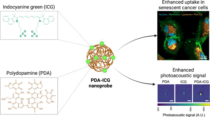
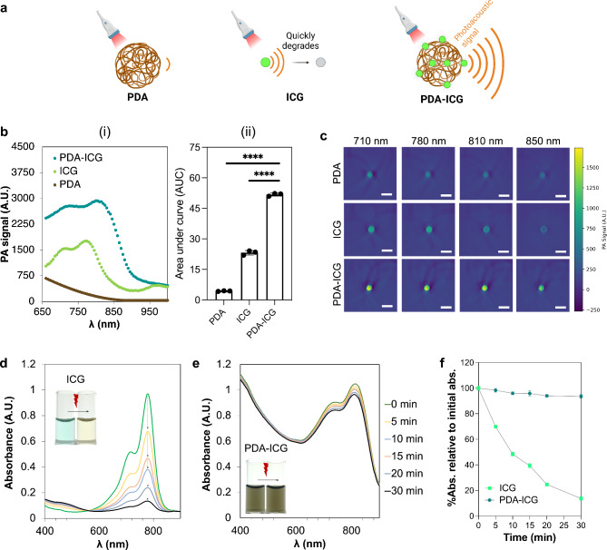
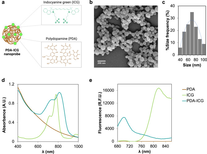
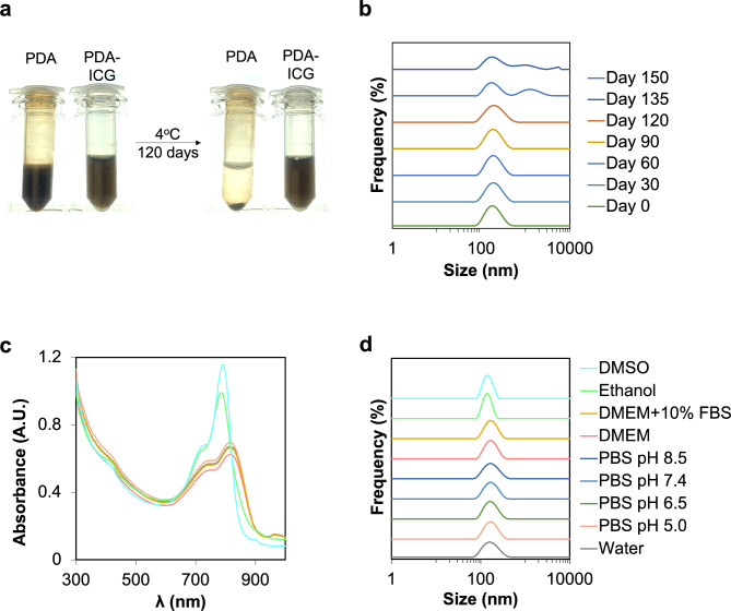
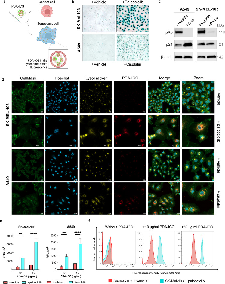
Cellular senescence is considered an important tumour suppression mechanism in response to damage and oncogenic stress in early lesions. However, when senescent cells are not immune-cleared and persist in the tumour microenvironment, they can drive a variety of tumour-promoting activities, including cancer initiation, progression, and metastasis. Additionally, there is compelling evidence demonstrating a direct connection between chemo(radio)therapy-induced senescence and the development of drug resistance and cancer recurrence. Therefore, detection of senescent cells in tissues holds great promise for predicting cancer occurrence earlier, assessing tumour progression, aiding patient stratification and prognosis, and informing about the efficacy of potential senotherapies. However, effective detection of senescent cells is limited by lack of biomarkers and readout strategies suitable for in vivo clinical imaging. To this end, a nanoprobe composed of biocompatible polydopamine (PDA) nanoparticle doped with FDA-approved indocyanine green (ICG) dye, namely PDA-ICG, was designed as a contrast agent for senescence detection using photoacoustic imaging (PAI). In an in vitro model of chemotherapy-induced senescence, PDA-ICG nanoprobe showed an elevated uptake in senescent cells relative to cancer cells. In addition to its improved photostability, 2.5-fold enhancement in photoacoustic signal relative to ICG was observed. Collectively, the results indicate that the PDA-ICG nanoprobe has the potential to be used as a contrast agent for senescence detection of chemotherapy-induced senescence using PAI.
细胞衰老被认为是一种重要的肿瘤抑制机制,可响应早期病变中的损伤和致癌应激。然而,当衰老细胞不能被免疫清除并在肿瘤微环境中持续存在时,它们可以驱动多种促进肿瘤的活动,包括癌症的发生、进展和转移。此外,有强有力的证据表明,化疗(放射)诱导的衰老与耐药性和癌症复发的发展之间存在直接联系。因此,在组织中检测衰老细胞有望更早地预测癌症发生,评估肿瘤进展,辅助患者分层和预后,并提供有关潜在衰老疗法疗效的信息。然而,衰老细胞的有效检测受到缺乏适合体内临床成像的生物标志物和读出策略的限制。为此,设计了一种由生物相容性聚多巴胺(PDA)纳米粒子掺杂 FDA 批准的吲哚菁绿(ICG)染料组成的纳米探针,即 PDA-ICG,用作光声成像(PAI)中衰老检测的对比剂。在化疗诱导的衰老体外模型中,PDA-ICG 纳米探针在衰老细胞中的摄取量相对于癌细胞有所增加。与 ICG 相比,光声信号增强了 2.5 倍。总的来说,这些结果表明,PDA-ICG 纳米探针有可能被用作 PAI 检测化疗诱导的衰老的对比剂。