Department of Interventional Radiology, Hospital Israelita Albert Einstein, São Paulo, SP, Brazil.
Department of Urology, Hospital Israelita Albert Einstein, São Paulo, SP, Brazil.
Einstein (Sao Paulo). 2024 Nov 22;22:eRC0779. doi: 10.31744/einstein_journal/2024RC0779. eCollection 2024.
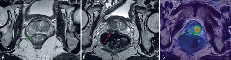
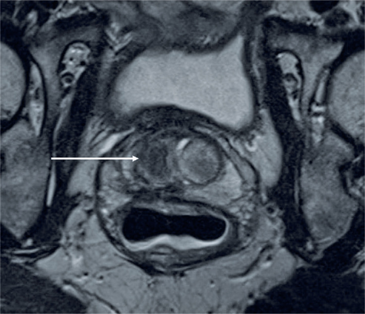
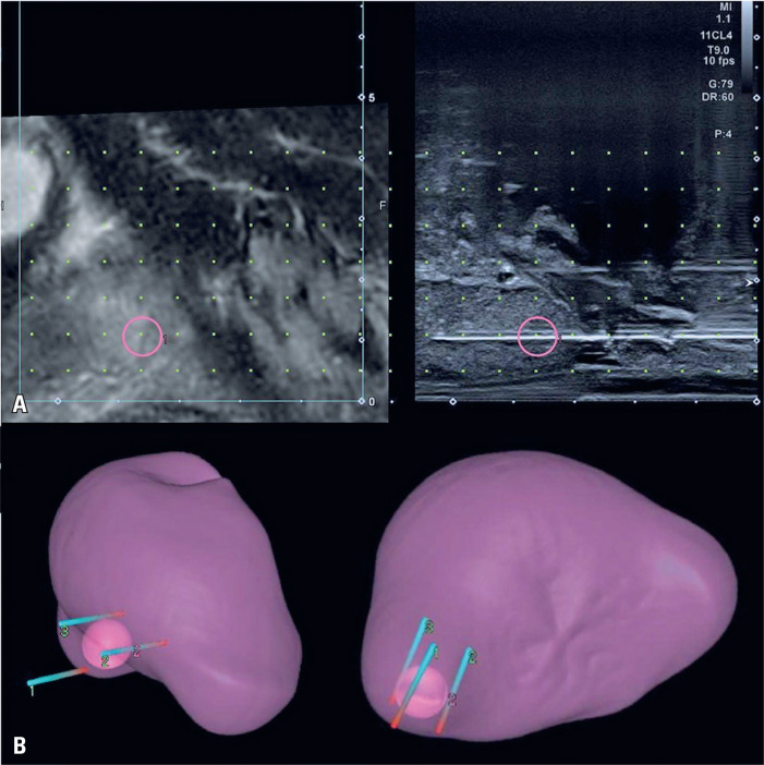
Radical treatment for prostate cancer is associated with significant morbidity. Percutaneous image-guided irreversible electroporation is a non-thermal ablative technique that has emerged as a valuable option. This study describes the case of a patient with prostate cancer who was successfully treated using irreversible electroporation. We report the case of a 72-year-old male patient who presented with elevated PSA (4.0ng/mL) during routine testing. Multiparametric magnetic resonance imaging of the prostate revealed a 0.8 cm lesion in the posterolateral aspect of the right midgland with marked hypointensity on ADC (ACR PI-RADS 4). The transperineal prostate revealed acinar adenocarcinoma (Gleason Score 3+3=6; International Society of Urological Pathology=1). Serum PSA levels reduced to 1.04ng/mL 32 days after the procedure and remained within normal limits (1.26ng/mL) after 349 days. Follow-up imaging performed 90 days later with prostate-specific membrane antigen PET/MRI showed size reduction, retraction, and diffuse hypointensity in the peripheral zone of the right prostate lobe, with no increase in prostate-specific membrane antigen uptake. Magnetic resonance imaging found no suspicious lesions 367 days after irreversible electroporation. At the final clinical follow-up at 390 days, the patient was asymptomatic. Our findings illustrate the potential of irreversible electroporation as a possible alternative treatment for prostate cancer.
根治性治疗前列腺癌与显著的发病率相关。经皮影像引导不可逆电穿孔是一种非热消融技术,已成为一种有价值的选择。本研究描述了一例成功接受不可逆电穿孔治疗的前列腺癌患者。我们报告了一例 72 岁男性患者的病例,该患者在常规检查中 PSA(4.0ng/mL)升高。前列腺多参数磁共振成像显示右中叶后外侧有 0.8cm 的病变,ADC 呈明显低信号(ACR PI-RADS 4)。经会阴前列腺显示腺泡性腺癌(Gleason 评分 3+3=6;国际泌尿病理学会=1)。手术后 32 天血清 PSA 水平降至 1.04ng/mL,349 天后仍在正常范围内(1.26ng/mL)。术后 90 天进行前列腺特异性膜抗原 PET/MRI 随访成像,显示右侧前列腺叶外周带体积缩小、回缩及弥漫性低信号,前列腺特异性膜抗原摄取未见增加。不可逆电穿孔 367 天后磁共振成像未发现可疑病变。在最终的 390 天临床随访时,患者无症状。我们的研究结果表明,不可逆电穿孔作为前列腺癌的一种可能替代治疗方法具有潜力。