Department of Orthopaedics, General Hospital of Central Theater Command, NO. 627, Wuluo Road, Hongshan District, Wuhan, Hubei Province, 430070, PR China.
The First Clinical Medical School of Southern Medical University, Guangzhou, Guangdong Province, PR China.
J Orthop Surg Res. 2024 Nov 28;19(1):808. doi: 10.1186/s13018-024-05305-7.
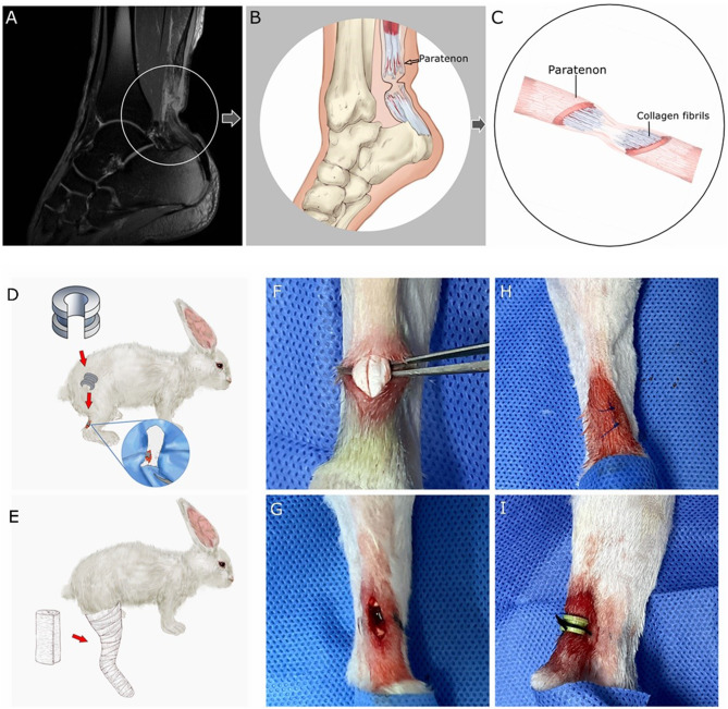
Limited research has focused on the correlation between an external compression and the regeneration of ruptured Achilles tendons. The aim of this study was to evaluate the influence of a constricted paratenon with external compression on the regeneration process of separated rabbit common calcanean tendon stumps.
A transection, establishing a 4 mm gap, was created in the right common calcanean tendon of 24 young adult male New Zealand white rabbits. The animals were assigned to two groups: In the control group, only received cast immobilization. In the constricted paratenon (CP) group, the rabbits had a local 3-dimensional printed clasp applied to mimic external compression and same cast immobilization as the control group. Morphologic, histologic and immunohistochemistry examinations were performed at 2 and 4 weeks postoperative.
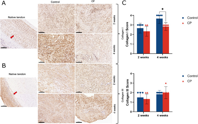
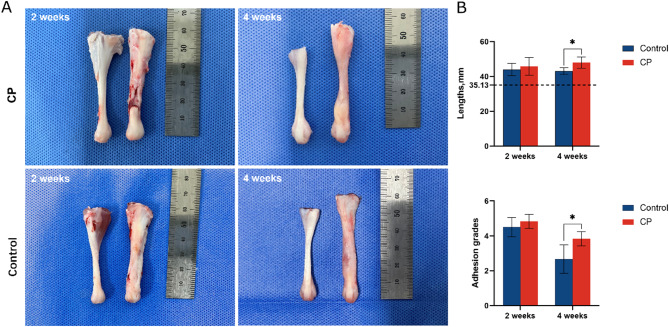
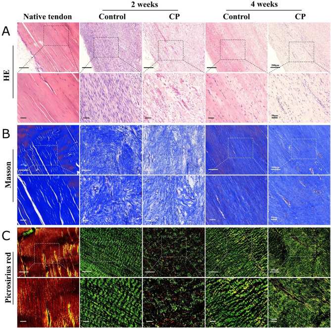
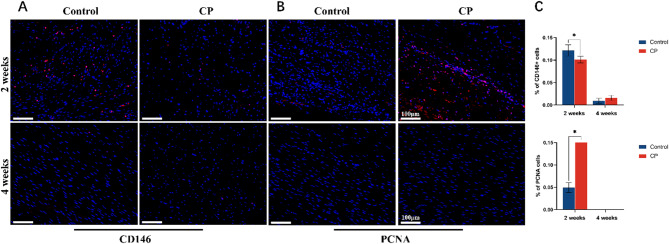
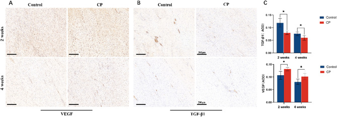
Separated tendon stumps were connected by novel granulated tendon fibrils in the control group. However, the regenerated tendon fibrils appeared insufficient in the CP group, the tendon length and the adhesion grade of the CP group was significantly larger than that of the control group at 4 weeks (P < 0.05, P = 0.030). Disorganized collagen and round-shaped fibroblasts were demonstrated in the CP group. A prolonged expression of proliferating cell nuclear antigen (PCNA) and lower intensity in clusters of differentiation 146 (CD146) were also shown in the CP group. A prolonged existence of the vascular endothelial growth factor (VEGF) and lesser intensity of the transforming growth factor-beta 1 (TGF-β1) were confirmed within this group. Furthermore, the CP group's expression had less collagen I than that of the control group at 4 weeks.
Sufficient regeneration can be obtained, even though there is an obvious gap between severed rabbit common calcanean tendon stumps. However, constricted paratenons with external compression can negatively influence the intrinsic regeneration process of the tendon fibrils and promotes the disorganization of regenerated collagen.
关于外部压迫与跟腱断裂后再生之间的相关性,目前的研究还很有限。本研究旨在评估外部压迫致缩窄性腱旁组织对兔跟腱断端再生过程的影响。
24 只新西兰大白兔右跟腱中后 1/3 处作横行切断,造成 4mm 间隙缺损。将动物随机分为两组:对照组仅接受石膏固定;缩窄性腱旁组织(CP)组,用 3D 打印扣环模拟外部压迫,同时接受石膏固定。术后 2 周和 4 周时进行形态学、组织学和免疫组织化学检查。
对照组断端之间有新生肉芽组织形成纤维束连接,但 CP 组再生的腱纤维束看起来不足,4 周时 CP 组的腱长度和粘连程度明显大于对照组(P<0.05,P=0.030)。CP 组胶原排列紊乱,成纤维细胞呈圆形。CP 组增殖细胞核抗原(PCNA)表达延长,簇分化 146(CD146)表达减弱。血管内皮生长因子(VEGF)持续存在,转化生长因子-β1(TGF-β1)表达减弱。此外,CP 组在第 4 周时的 I 型胶原表达也低于对照组。
即使兔跟腱断端之间存在明显的间隙,也能获得充分的再生。然而,外部压迫致缩窄性腱旁组织可对腱纤维的内在再生过程产生负面影响,并促进胶原的排列紊乱。