Suresh Samyuktha, Karthik Gayathri, Ouyang John F, Chrysostomou Vicki, Tang See Aik, Petretto Enrico, Crowston Jonathan G, Bell Katharina C
Neuroscience and Behavioural Diseases and Eye-ACP, Centre for Vision Research, Duke-NUS Medical School, Singapore, Singapore.
Programme in Cardiovascular and Metabolic Disorders, Duke-NUS Medical School, Singapore, Singapore.
NPJ Aging. 2024 Nov 30;10(1):60. doi: 10.1038/s41514-024-00187-9.
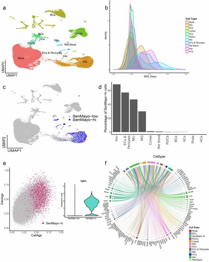
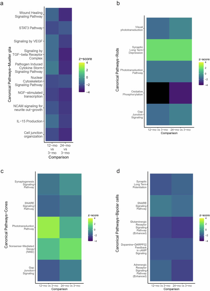
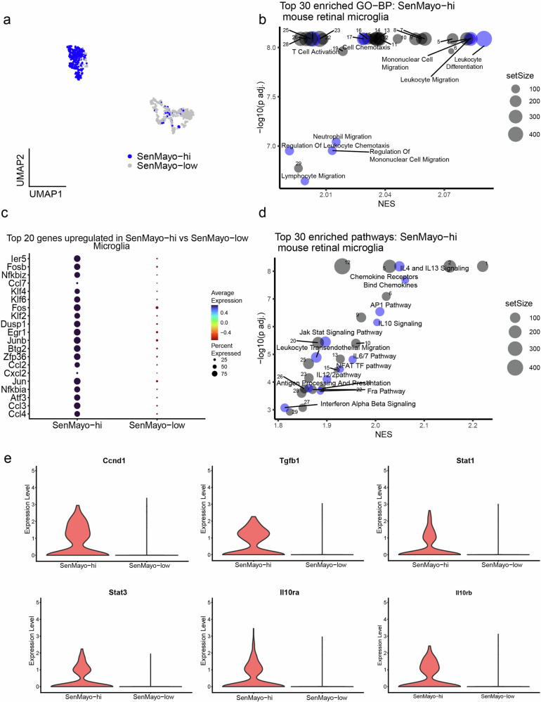
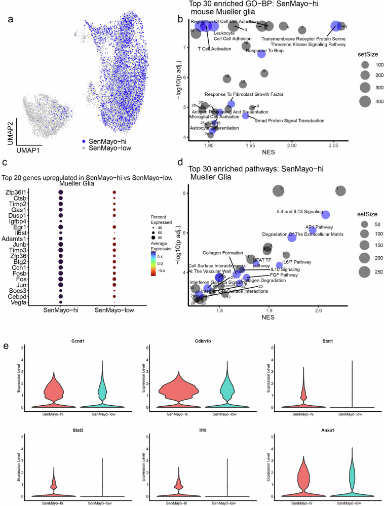
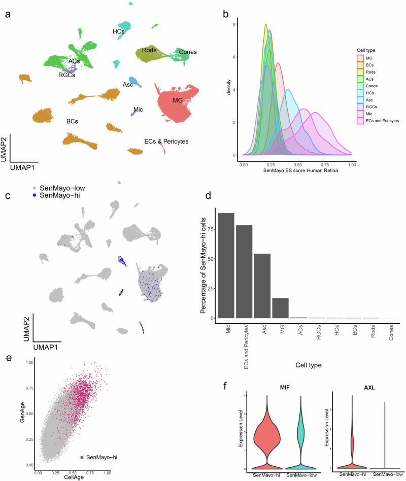
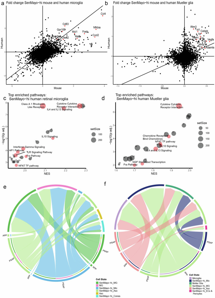
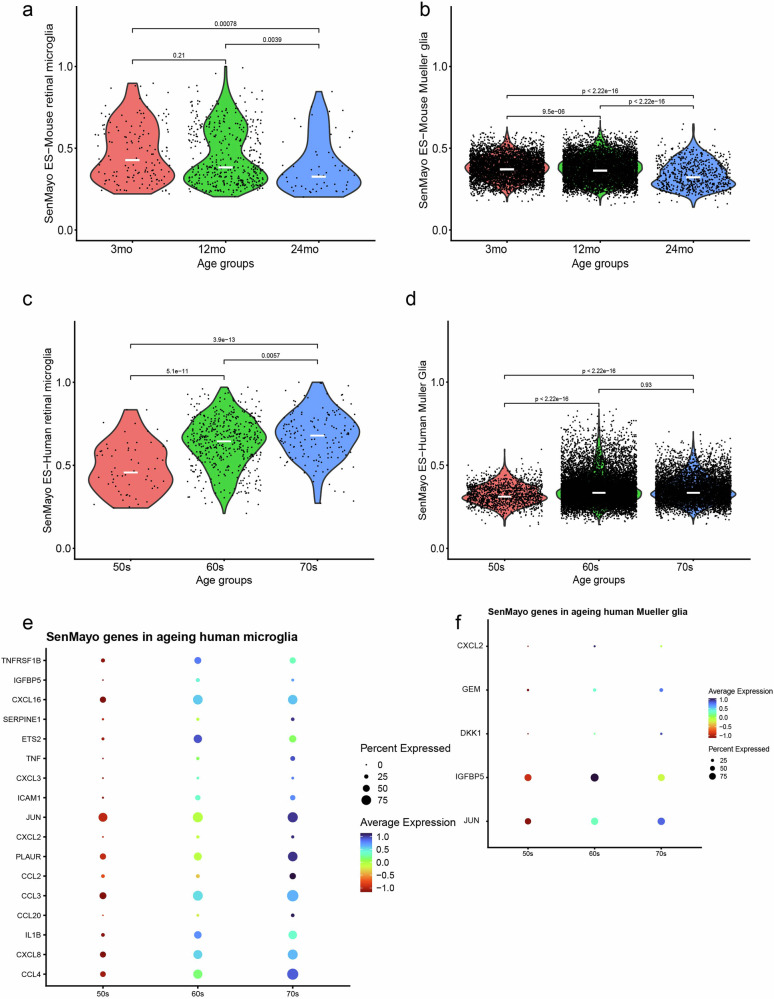
There is a growing need to better characterise senescent cells in the CNS and retina. The recently published SenMayo gene panel was developed to identify transcriptomic signatures of senescence across multiple organ systems, but the retina was not included. While other approaches have identified senescent signatures in the retina, these have largely focused on experimental models in young animals. We therefore conducted a detailed single-cell RNA-seq analysis to identify senescent cell populations in the retina of different aged mice and compared these with five comprehensive human and mouse retina and brain transcriptome datasets. Transcriptomic signatures of senescence were most apparent in mouse and human retinal glial cells, with IL4, 13 and 10 and the AP1 pathway being the most prominent markers involved. Similar levels of transcriptional senescence were observed in the retinal glia of young and old mice, whereas the human retina showed significantly increased enrichment scores with advancing age.
更好地表征中枢神经系统和视网膜中的衰老细胞的需求日益增长。最近发布的SenMayo基因面板旨在识别多个器官系统中衰老的转录组特征,但未包括视网膜。虽然其他方法已在视网膜中识别出衰老特征,但这些方法主要集中在幼小动物的实验模型上。因此,我们进行了详细的单细胞RNA测序分析,以识别不同年龄小鼠视网膜中的衰老细胞群体,并将其与五个全面的人类和小鼠视网膜及脑转录组数据集进行比较。衰老的转录组特征在小鼠和人类视网膜神经胶质细胞中最为明显,其中白细胞介素4、13和10以及AP1信号通路是最突出的相关标志物。在年轻和老年小鼠的视网膜神经胶质细胞中观察到相似水平的转录衰老,而人类视网膜随着年龄的增长显示出显著增加的富集分数。