Department of Biology, Animal Physiology and Neurobiology Division, Neural Circuit Development & Regeneration Research Group, KU Leuven, Leuven Brain Institute, Leuven, Belgium.
University College London, Institute of Ophthalmology, London, UK.
Aging Cell. 2024 Aug;23(8):e14192. doi: 10.1111/acel.14192. Epub 2024 May 14.
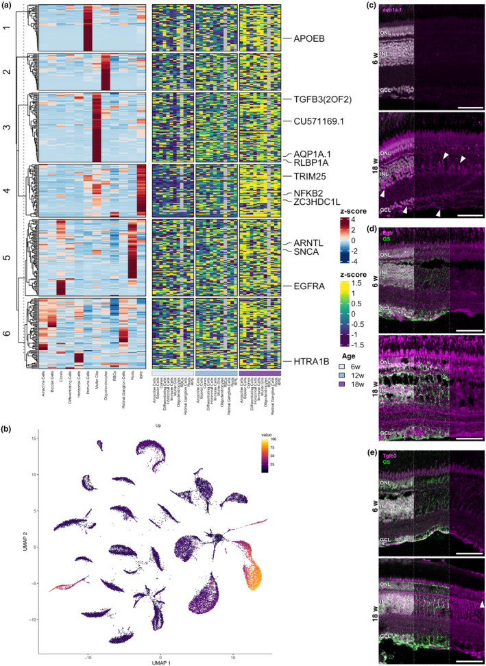
Age-related vision loss caused by retinal neurodegenerative pathologies is becoming more prevalent in our ageing society. To understand the physiological and molecular impact of ageing on retinal homeostasis, we used the short-lived African turquoise killifish, a model known to naturally develop central nervous system (CNS) ageing hallmarks and vision loss. Bulk and single-cell RNA-sequencing (scRNAseq) of three age groups (6-, 12-, and 18-week-old) identified transcriptional ageing fingerprints in the killifish retina, unveiling pathways also identified in the aged brain, including oxidative stress, gliosis, and inflammageing. These findings were comparable to observations in the ageing mouse retina. Additionally, transcriptional changes in genes related to retinal diseases, such as glaucoma and age-related macular degeneration, were observed. The cellular heterogeneity in the killifish retina was characterized, confirming the presence of all typical vertebrate retinal cell types. Data integration from age-matched samples between the bulk and scRNAseq experiments revealed a loss of cellular specificity in gene expression upon ageing, suggesting potential disruption in transcriptional homeostasis. Differential expression analysis within the identified cell types highlighted the role of glial/immune cells as important stress regulators during ageing. Our work emphasizes the value of the fast-ageing killifish in elucidating molecular signatures in age-associated retinal disease and vision decline. This study contributes to the understanding of how age-related changes in molecular pathways may impact CNS health, providing insights that may inform future therapeutic strategies for age-related pathologies.
年龄相关性视网膜神经退行性病变导致的视力丧失在我们老龄化的社会中越来越普遍。为了了解衰老对视网膜内稳态的生理和分子影响,我们使用了短寿命的非洲绿松石食蚊鱼,这种鱼类被认为会自然发展出中枢神经系统(CNS)衰老特征和视力丧失。对三个年龄段(6 周、12 周和 18 周)的食蚊鱼视网膜进行 bulk 和单细胞 RNA 测序(scRNAseq),确定了食蚊鱼视网膜中的转录衰老特征,揭示了在衰老大脑中也发现的途径,包括氧化应激、神经胶质增生和炎症衰老。这些发现与衰老小鼠视网膜的观察结果相当。此外,还观察到与视网膜疾病(如青光眼和年龄相关性黄斑变性)相关的基因的转录变化。对食蚊鱼视网膜的细胞异质性进行了特征描述,证实了所有典型的脊椎动物视网膜细胞类型的存在。对 bulk 和 scRNAseq 实验之间年龄匹配样本的数据进行整合,揭示了衰老时基因表达的细胞特异性丧失,表明转录内稳态可能受到潜在破坏。在鉴定出的细胞类型内进行差异表达分析,强调了神经胶质/免疫细胞在衰老过程中作为重要应激调节剂的作用。我们的工作强调了快速衰老的食蚊鱼在阐明与年龄相关的视网膜疾病和视力下降的分子特征方面的价值。这项研究有助于了解分子途径的年龄相关性变化如何影响中枢神经系统健康,为针对与年龄相关的病理学的未来治疗策略提供了见解。