• 文献检索
  • 文档翻译
  • 深度研究
  • 学术资讯
  • Suppr Zotero 插件Zotero 插件
  • 邀请有礼
  • 套餐&价格
  • 历史记录
应用&插件
Suppr Zotero 插件Zotero 插件浏览器插件Mac 客户端Windows 客户端微信小程序
定价
高级版会员购买积分包购买API积分包
服务
文献检索文档翻译深度研究API 文档MCP 服务
关于我们
关于 Suppr公司介绍联系我们用户协议隐私条款
关注我们

Suppr 超能文献

核心技术专利:CN118964589B侵权必究
粤ICP备2023148730 号-1Suppr @ 2026

文献检索

告别复杂PubMed语法,用中文像聊天一样搜索,搜遍4000万医学文献。AI智能推荐,让科研检索更轻松。

立即免费搜索

文件翻译

保留排版,准确专业,支持PDF/Word/PPT等文件格式,支持 12+语言互译。

免费翻译文档

深度研究

AI帮你快速写综述,25分钟生成高质量综述,智能提取关键信息,辅助科研写作。

立即免费体验

早期青光眼黄斑神经节细胞层厚度及灌注密度变化的序列与可检测性

Sequence and Detectability of Changes in Macular Ganglion Cell Layer Thickness and Perfusion Density in Early Glaucoma.

作者信息

Tomita Ryo, Smith Corey A, Dyachok Oksana M, Sharpe Glen P, Rafuse Paul E, Shuba Lesya M, Nicolela Marcelo T, Chauhan Balwantray C

机构信息

Department of Ophthalmology and Visual Sciences, Dalhousie University, Halifax, Nova Scotia, Canada.

出版信息

Invest Ophthalmol Vis Sci. 2024 Dec 2;65(14):8. doi: 10.1167/iovs.65.14.8.

DOI:10.1167/iovs.65.14.8
PMID:39625439
原文链接:https://pmc.ncbi.nlm.nih.gov/articles/PMC11627249/
Abstract

PURPOSE

To investigate whether macular perfusion density (PD) changes measured over time by optical coherence tomography angiography (OCTA) are detectable before progressive macular ganglion cell layer (GCL) thinning in early glaucoma.

METHODS

This prospective longitudinal cohort study involved patients with early open-angle glaucoma and healthy subjects imaged by OCT and OCTA every 4 months. GCL thickness and macular PD were evaluated in 16 tiles in the macula. We estimated baseline percentage losses of GCL thickness or macular PD in glaucoma patients with age-corrected normative values derived from the healthy subjects. Additionally, the threshold slope separating glaucoma patients from healthy subjects with 90% specificity was used to determine the number of patients with steeper slopes than the threshold slope.

RESULTS

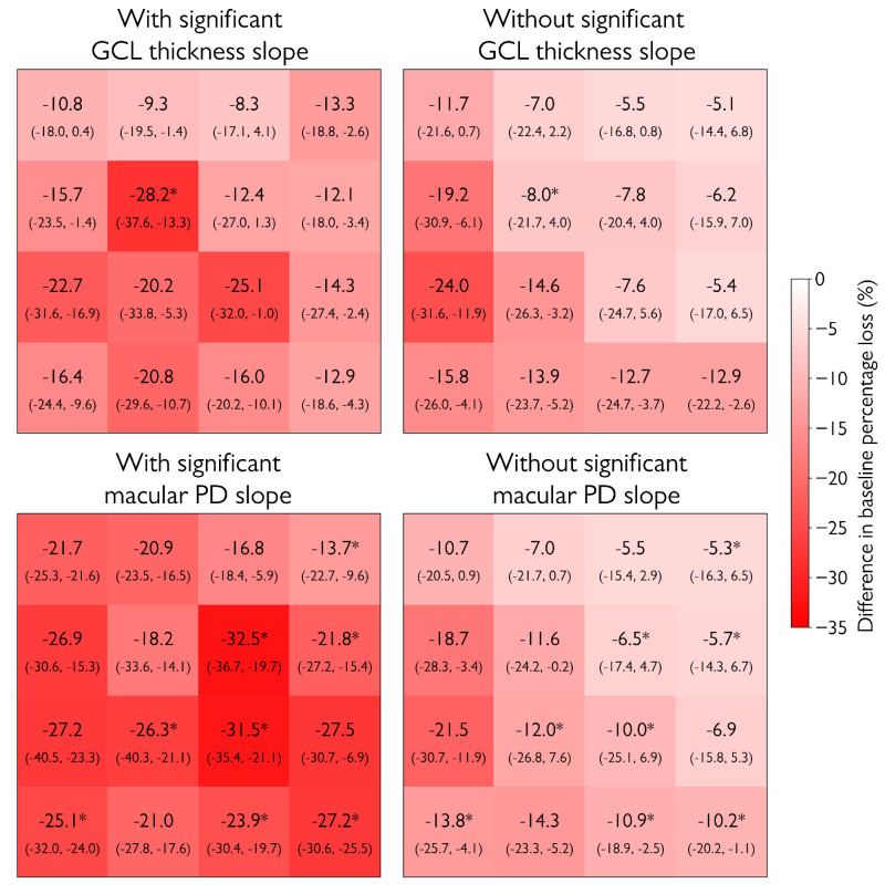
Eighty patients with glaucoma and 42 healthy subjects were included. In eight tiles (50%), patients with a significant macular PD slope had a significantly greater baseline percentage loss of GCL thickness relative to macular PD compared to patients without a significant macular PD slope. Furthermore, in 15 tiles (94%), a greater baseline percentage loss of GCL thickness relative to PD was significantly correlated with faster PD slopes. In contrast, only one tile (6%) showed these significant trends for GCL slopes. The number of patients with faster GCL slopes than threshold slopes was significantly larger than patients with faster PD slopes in 12 tiles (75%).

CONCLUSIONS

A decrease in GCL thickness precedes a measurable decrease in macular PD. Early glaucomatous progression is more frequently detectable with changes in GCL thickness compared to macular PD.

摘要

目的

研究在早期青光眼患者黄斑神经节细胞层(GCL)逐渐变薄之前,通过光学相干断层扫描血管造影(OCTA)随时间测量的黄斑灌注密度(PD)变化是否可被检测到。

方法

这项前瞻性纵向队列研究纳入了早期开角型青光眼患者和健康受试者,每4个月进行一次OCT和OCTA成像。对黄斑区的16个区域评估GCL厚度和黄斑PD。我们根据健康受试者的年龄校正规范值估计青光眼患者GCL厚度或黄斑PD的基线百分比损失。此外,使用将青光眼患者与具有90%特异性的健康受试者区分开的阈值斜率来确定斜率比阈值斜率更陡的患者数量。

结果

纳入了80例青光眼患者和42例健康受试者。在8个区域(50%)中,与没有显著黄斑PD斜率的患者相比,具有显著黄斑PD斜率的患者GCL厚度相对于黄斑PD的基线百分比损失显著更大。此外,在15个区域(94%)中,GCL厚度相对于PD的基线百分比损失更大与更快的PD斜率显著相关。相比之下,只有1个区域(6%)显示出GCL斜率的这些显著趋势。在12个区域(75%)中,GCL斜率比阈值斜率更快的患者数量显著多于PD斜率更快的患者。

结论

GCL厚度的降低先于黄斑PD的可测量降低。与黄斑PD相比,早期青光眼进展更常通过GCL厚度的变化被检测到。

https://cdn.ncbi.nlm.nih.gov/pmc/blobs/2d8b/11627249/c159d9457e39/iovs-65-14-8-f006.jpg
https://cdn.ncbi.nlm.nih.gov/pmc/blobs/2d8b/11627249/b87b5a343cfd/iovs-65-14-8-f001.jpg
https://cdn.ncbi.nlm.nih.gov/pmc/blobs/2d8b/11627249/b07a3c257617/iovs-65-14-8-f002.jpg
https://cdn.ncbi.nlm.nih.gov/pmc/blobs/2d8b/11627249/7d771d0ca038/iovs-65-14-8-f003.jpg
https://cdn.ncbi.nlm.nih.gov/pmc/blobs/2d8b/11627249/6643068b2807/iovs-65-14-8-f004.jpg
https://cdn.ncbi.nlm.nih.gov/pmc/blobs/2d8b/11627249/cd693d41d5aa/iovs-65-14-8-f005.jpg
https://cdn.ncbi.nlm.nih.gov/pmc/blobs/2d8b/11627249/c159d9457e39/iovs-65-14-8-f006.jpg

相似文献

1
Sequence and Detectability of Changes in Macular Ganglion Cell Layer Thickness and Perfusion Density in Early Glaucoma.早期青光眼黄斑神经节细胞层厚度及灌注密度变化的序列与可检测性
Invest Ophthalmol Vis Sci. 2024 Dec 2;65(14):8. doi: 10.1167/iovs.65.14.8.
2
Ganglion Cell Complex Thickness and Macular Vessel Density Loss in Primary Open-Angle Glaucoma.原发性开角型青光眼的神经节细胞复合体厚度和黄斑血管密度损失。
Ophthalmology. 2020 Aug;127(8):1043-1052. doi: 10.1016/j.ophtha.2019.12.030. Epub 2020 Jan 13.
3
Discrepancy in Loss of Macular Perfusion Density and Ganglion Cell Layer Thickness in Early Glaucoma.早期青光眼黄斑血流密度与神经节细胞层厚度的差异。
Am J Ophthalmol. 2021 Jan;221:39-47. doi: 10.1016/j.ajo.2020.08.031. Epub 2020 Aug 20.
4
Comparative evaluation of OCT with OCTA changes at the optic disc and macula in glaucoma suspect and early glaucoma.青光眼可疑患者和早期青光眼患者视盘和黄斑区光学相干断层扫描(OCT)与光学相干断层扫描血管造影(OCTA)变化的比较评估
Indian J Ophthalmol. 2025 Mar 1;73(Suppl 2):S260-S266. doi: 10.4103/IJO.IJO_2575_23. Epub 2024 Oct 25.
5
Evaluation of Layer-by-Layer Segmented Ganglion Cell Complex Thickness for Detecting Early Glaucoma According to Different Macular Grids.根据不同黄斑网格评估逐层分割的神经节细胞复合体厚度以检测早期青光眼
J Glaucoma. 2017 Aug;26(8):712-717. doi: 10.1097/IJG.0000000000000709.
6
Ganglion Cell-Inner Plexiform Layer Thickness in Different Glaucoma Stages Measured by Optical Coherence Tomography.光学相干断层扫描测量不同青光眼阶段的神经节细胞-内网状层厚度
Ophthalmic Res. 2018;59(3):148-154. doi: 10.1159/000478052. Epub 2017 Sep 7.
7
Glaucomatous Focal Perfusion Loss in the Macula Measured by Optical Coherence Tomographic Angiography.光学相干断层扫描血管造影测量的黄斑区青光眼局灶性灌注损失。
Am J Ophthalmol. 2024 Dec;268:181-189. doi: 10.1016/j.ajo.2024.07.008. Epub 2024 Jul 14.
8
Longitudinal Macular Structure-Function Relationships in Glaucoma.青光眼的纵向黄斑结构-功能关系。
Ophthalmology. 2020 Jul;127(7):888-900. doi: 10.1016/j.ophtha.2020.01.023. Epub 2020 Jan 22.
9
Differences in swept-source OCT angiography of the macular capillary network in high tension and normal tension glaucoma.高眼压症和正常眼压青光眼的黄斑毛细血管网络扫频源光学相干断层扫描血管造影的差异。
Curr Eye Res. 2020 Sep;45(9):1168-1172. doi: 10.1080/02713683.2020.1722178. Epub 2020 Feb 3.
10
Projection-Resolved Optical Coherence Tomography Angiography of Macular Retinal Circulation in Glaucoma.青光眼黄斑视网膜循环的投影分辨光学相干断层扫描血管造影
Ophthalmology. 2017 Nov;124(11):1589-1599. doi: 10.1016/j.ophtha.2017.06.002. Epub 2017 Jul 1.

本文引用的文献

1
Variability of scan quality and perfusion density in longitudinal optical coherence tomography angiography imaging.纵向光学相干断层扫描血管造影成像中的扫描质量和灌注密度的可变性。
Br J Ophthalmol. 2024 Jun 20;108(7):978-983. doi: 10.1136/bjo-2022-322979.
2
Progressive Macular Vessel Density Loss and Visual Field Progression in Open-angle Glaucoma Eyes with Central Visual Field Damage.中心视野损伤的开角型青光眼眼中进行性黄斑血管密度损失和视野进展。
Ophthalmol Glaucoma. 2024 Jan-Feb;7(1):16-29. doi: 10.1016/j.ogla.2023.06.009. Epub 2023 Jun 26.
3
Evaluation of the long-term variability of macular OCT/OCTA and visual field parameters.
黄斑OCT/OCTA及视野参数的长期变异性评估
Br J Ophthalmol. 2024 Jan 29;108(2):211-216. doi: 10.1136/bjo-2022-322470.
4
Quantitative Parameters from OCT Angiography in Patients with Diabetic Retinopathy and in Those with Only Peripheral Retinopathy Compared with Control Participants.糖尿病视网膜病变患者、仅患有周边视网膜病变患者与对照参与者相比,光学相干断层扫描血管造影的定量参数。
Ophthalmol Sci. 2021 May 26;1(2):100030. doi: 10.1016/j.xops.2021.100030. eCollection 2021 Jun.
5
Impact of Glaucoma Severity on Rates of Neuroretinal Rim, Retinal Nerve Fiber Layer, and Macular Ganglion Cell Layer Thickness Change.青光眼严重程度对视神经视网膜边缘、视网膜神经纤维层和黄斑神经节细胞层厚度变化率的影响。
Am J Ophthalmol. 2022 Jul;239:115-121. doi: 10.1016/j.ajo.2022.01.019. Epub 2022 Feb 2.
6
Long-term reproducibility of optical coherence tomography angiography in healthy and stable glaucomatous eyes.健康及稳定青光眼眼中光学相干断层扫描血管造影的长期可重复性。
Br J Ophthalmol. 2023 May;107(5):657-662. doi: 10.1136/bjophthalmol-2021-320034. Epub 2021 Dec 21.
7
Demographic, Comorbid, and Clinical Variables Associated With Pointwise Visual Field Damage in Glaucoma: Data From the AGIS and CIGTS Clinical Trials.与青光眼点状视野损伤相关的人口统计学、合并症和临床变量:来自 AGIS 和 CIGTS 临床试验的数据。
Transl Vis Sci Technol. 2021 Oct 4;10(12):28. doi: 10.1167/tvst.10.12.28.
8
Vascular biomarkers from optical coherence tomography angiography and glaucoma: where do we stand in 2021?光学相干断层扫描血管造影和青光眼的血管生物标志物:2021 年我们处于什么位置?
Acta Ophthalmol. 2022 Mar;100(2):e377-e385. doi: 10.1111/aos.14982. Epub 2021 Aug 6.
9
Macular Thickness and Microvasculature Loss in Glaucoma Suspect Eyes.疑似青光眼患者眼部的黄斑厚度及微血管损失
Ophthalmol Glaucoma. 2022 Mar-Apr;5(2):170-178. doi: 10.1016/j.ogla.2021.07.009. Epub 2021 Jul 30.
10
OCT Angiography Artifacts in Glaucoma.青光眼的 OCT 血管造影伪影。
Ophthalmology. 2021 Oct;128(10):1426-1437. doi: 10.1016/j.ophtha.2021.03.036. Epub 2021 Apr 2.