Saitou Toshinori, Aikawa Tadao, Manabe Osamu, Fujimoto Shinichiro, Matsue Yuya, Nagase Atsushi, Toyama Hiroaki, Kudo Tamaki, Oyama-Manabe Noriko, Minamino Tohru
Department of Radiology, Hokkaido Cardiovascular Hospital, Hokkaido, Japan.
Department of Cardiovascular Biology and Medicine, Juntendo University Graduate School of Medicine, Tokyo, Japan.
Ann Nucl Cardiol. 2024;10(1):29-37. doi: 10.17996/anc.24-00002. Epub 2024 Oct 31.
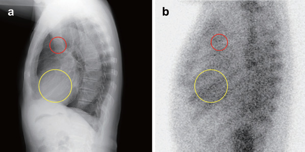
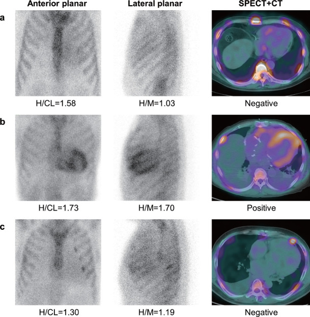
: Lateral planar Tc-pyrophosphate (PYP) imaging is recommended as a standardized acquisition method because it helps separate extracardiac uptake from the myocardium. We evaluated its discriminatory performance in detecting myocardial PYP uptake, using single-photon emission computed tomography (SPECT) as a reference standard. : We retrospectively evaluated 170 patients who underwent PYP imaging for suspected transthyretin cardiac amyloidosis. Anterior and lateral planar imaging and SPECT were performed 3 h after PYP administration. The myocardial PYP uptake on planar and SPECT images was visually assessed and quantified using the heart-to-contralateral lung uptake (H/CL) ratio. The heart-to-mediastinum uptake (H/M) ratio was calculated as the mean count of the region of interest in the heart divided by that in the superior mediastinum on lateral planar images. : Patients with PYP SPECT-positive results had significantly higher H/M ratios at 3 h than those with PYP SPECT-negative results (1.23 [interquartile range: IQR, 1.15-1.43] vs. 1.08 [IQR, 1.02-1.16]; <0.001). A reclassification analysis that added the H/M ratio to visual scores for detecting positive PYP SPECT yielded a significant improvement with a net reclassification improvement (NRI) of 0.56 (95%CI, 0.25-0.87; <0.001) and integrated discrimination improvement (IDI) of 0.038 (95%CI, 0.005-0.072; =0.026). The H/M ratio significantly improved the predictive ability of SPECT findings based on the visual scores and H/CL ratio with an NRI of 0.49 (95%CI, 0.18-0.81; =0.003) and IDI of 0.036 (95%CI, 0.004-0.069; =0.029). : Adding the H/M ratio derived from lateral planar PYP imaging to visual scores or the H/CL ratio on anterior planar images improved the accuracy of detecting significant myocardial uptake on SPECT.
推荐采用平面侧位锝焦磷酸盐(PYP)显像作为标准化采集方法,因为它有助于区分心肌摄取与心外摄取。我们以单光子发射计算机断层扫描(SPECT)作为参考标准,评估了其在检测心肌PYP摄取方面的鉴别性能。
我们回顾性评估了170例因疑似转甲状腺素蛋白心脏淀粉样变性而接受PYP显像的患者。在注射PYP后3小时进行前位和平面侧位显像以及SPECT检查。通过心脏与对侧肺摄取(H/CL)比值对平面和SPECT图像上的心肌PYP摄取进行视觉评估和定量分析。心脏与纵隔摄取(H/M)比值计算为平面侧位图像上心感兴趣区的平均计数除以上纵隔感兴趣区的平均计数。
PYP SPECT检查结果为阳性的患者在3小时时的H/M比值显著高于PYP SPECT检查结果为阴性的患者(1.23[四分位间距:IQR,1.15 - 1.43] 对比 1.08[IQR,1.02 - 1.16];P<0.001)。将H/M比值添加到用于检测PYP SPECT阳性的视觉评分中的重新分类分析显示有显著改善,净重新分类改善(NRI)为0.56(95%CI,0.25 - 0.87;P<0.001),综合鉴别改善(IDI)为0.038(95%CI,0.005 - 0.072;P = 0.026)。基于视觉评分和H/CL比值,H/M比值显著提高了SPECT检查结果的预测能力,NRI为0.49(95%CI,0.18 - 0.81;P = 0.003),IDI为0.036(95%CI,0.004 - 0.069;P = 0.029)。
将平面侧位PYP显像得出的H/M比值添加到视觉评分或前位平面图像的H/CL比值中,可提高检测SPECT上显著心肌摄取的准确性。