• 文献检索
  • 文档翻译
  • 深度研究
  • 学术资讯
  • Suppr Zotero 插件Zotero 插件
  • 邀请有礼
  • 套餐&价格
  • 历史记录
应用&插件
Suppr Zotero 插件Zotero 插件浏览器插件Mac 客户端Windows 客户端微信小程序
定价
高级版会员购买积分包购买API积分包
服务
文献检索文档翻译深度研究API 文档MCP 服务
关于我们
关于 Suppr公司介绍联系我们用户协议隐私条款
关注我们

Suppr 超能文献

核心技术专利:CN118964589B侵权必究
粤ICP备2023148730 号-1Suppr @ 2026

文献检索

告别复杂PubMed语法,用中文像聊天一样搜索,搜遍4000万医学文献。AI智能推荐,让科研检索更轻松。

立即免费搜索

文件翻译

保留排版,准确专业,支持PDF/Word/PPT等文件格式,支持 12+语言互译。

免费翻译文档

深度研究

AI帮你快速写综述,25分钟生成高质量综述,智能提取关键信息,辅助科研写作。

立即免费体验

局灶性肝病变:通过超分辨率超声成像进行多参数微血管特征分析

Focal liver lesions: multiparametric microvasculature characterization via super-resolution ultrasound imaging.

作者信息

Zeng Qian-Qian, An Shi-Zhe, Chen Chao-Nan, Wang Zhen, Liu Jia-Cheng, Wan Ming-Xi, Zong Yu-Jin, Jian Xiao-Hua, Yu Jie, Liang Ping

机构信息

Department of Interventional Ultrasound, Senior Department of Oncology, The Fifth Medical Center of PLA General Hospital, Fengtai District, Beijing, 100853, China.

Chinese People's Liberation Army (PLA) Medical School, Haidian District, Beijing, 100853, China.

出版信息

Eur Radiol Exp. 2024 Dec 5;8(1):138. doi: 10.1186/s41747-024-00540-3.

DOI:10.1186/s41747-024-00540-3
PMID:39636384
原文链接:https://pmc.ncbi.nlm.nih.gov/articles/PMC11621259/
Abstract

BACKGROUND

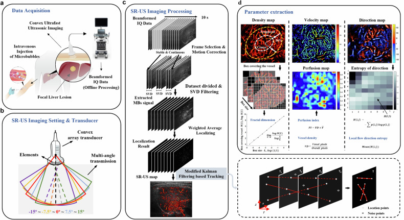
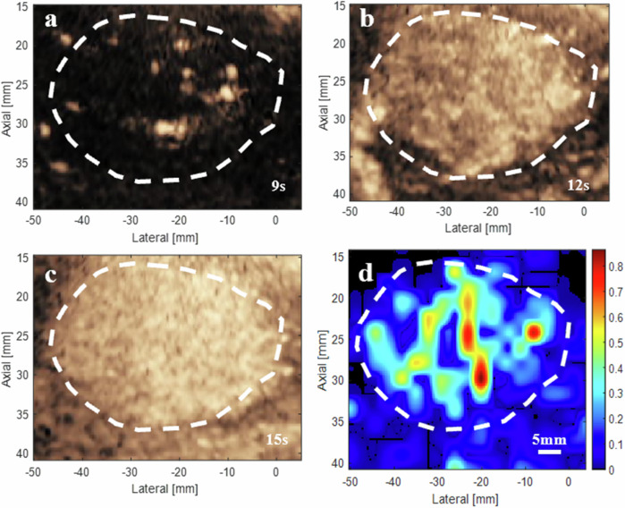
Noninvasive and functional imaging of the focal liver lesion (FLL) vasculature at microscopic scales is clinically challenging. We investigated the feasibility of using super-resolution ultrasound (SR-US) imaging for visualizing and quantifying the microvasculature of intraparenchymal FLLs.

METHODS

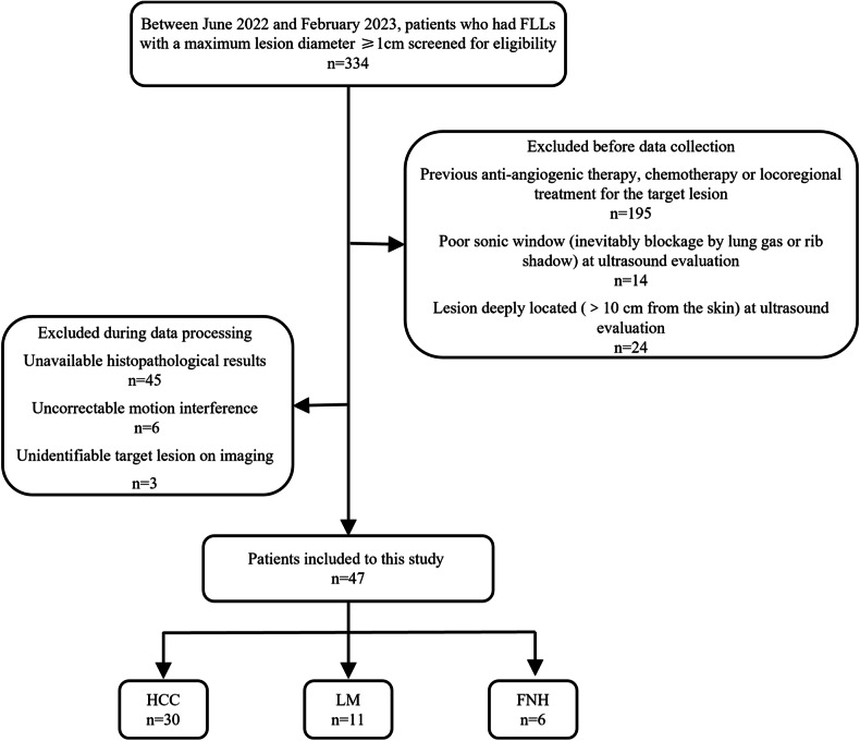
Patients with FLLs between June 2022 and February 2023 were prospectively screened. Following bolus injection of microbubbles at clinical concentration, SR-US was performed using a high frame rate (350-500 Hz) modified ultrasound scanner and a convex array transducer with a central frequency of 3.1 MHz.

RESULTS

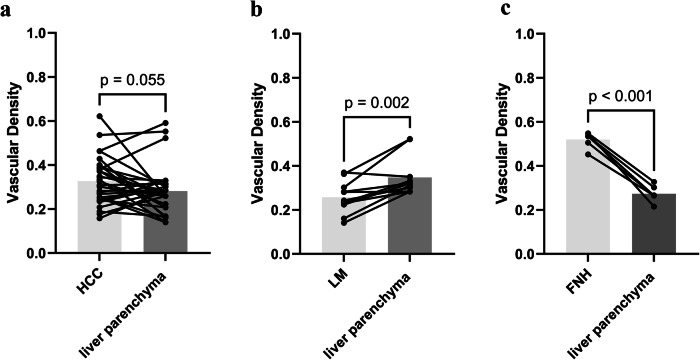
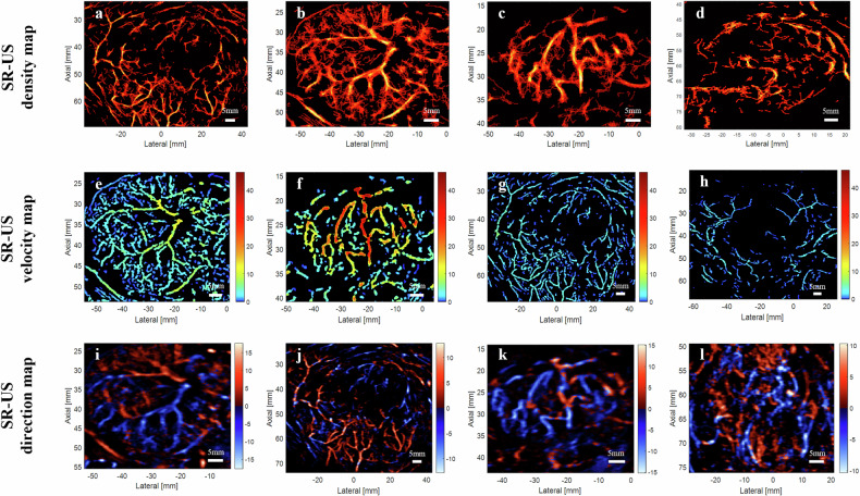
In total, 47 pathologically proven FLLs at a depth of 5.7 ± 1.7 cm (mean ± standard deviation) were included: 30 hepatocellular carcinomas (HCC), 11 liver metastases (LM), and 6 focal nodular hyperplasias (FNH). The smallest detectable vessel size of the hepatic microvasculature was 128.4 ± 18.6 μm (mean ± standard deviation) at a depth of 8 cm. Significant differences were observed among the three types of lesions in terms of pattern categories, vessel density, minimum flow velocity, and perfusion index. We observed higher vessel density for FNH versus liver parenchyma (p < 0.001) as well as fractal dimension and local flow direction entropy value for FNH versus HCC (p = 0.002 and p < 0.001, respectively) and for FNH versus LM (p = 0.006 and p = 0.002, respectively).

CONCLUSION

Multiparametric SR-US showed that these three pathological types of FLLs have specific microvascular phenotypes. Vessel density, fractal dimension and local flow direction entropy served as valuable parameters in distinguishing between benign and malignant FLLs.

TRIAL REGISTRATION

ClinicalTrials.gov (NCT06018142).

RELEVANCE STATEMENT

Multiparametric SR-US imaging offers precise morphological and functional assessment of the microvasculature of intraparenchymal focal liver lesions, providing insights into tumor heterogeneity and angiogenesis.

KEY POINTS

Super-resolution (SR)-US imaging allowed morphological and functional evaluation of intraparenchymal hepatic lesion microvasculature. Hepatocellular carcinoma, liver metastasis, and focal nodular hyperplasia exhibit distinct microvascular architectures and hemodynamic profiles. Multiparametric microvasculature characterization via SR-US imaging facilitates the differentiation between benign and malignant microvascular phenotypes.

摘要

背景

在微观尺度上对肝脏局灶性病变(FLL)的血管系统进行无创性和功能性成像在临床上具有挑战性。我们研究了使用超分辨率超声(SR-US)成像来可视化和量化肝实质内FLL微血管系统的可行性。

方法

对2022年6月至2023年2月期间患有FLL的患者进行前瞻性筛查。在以临床浓度推注微泡后,使用高帧率(350 - 500Hz)的改良超声扫描仪和中心频率为3.1MHz的凸阵换能器进行SR-US检查。

结果

总共纳入了47个经病理证实的FLL,深度为5.7±1.7cm(平均值±标准差),其中包括30例肝细胞癌(HCC)、11例肝转移瘤(LM)和6例局灶性结节性增生(FNH)。在8cm深度处,肝脏微血管系统的最小可检测血管大小为128.4±18.6μm(平均值±标准差)。在模式类别、血管密度、最小流速和灌注指数方面,三种类型的病变之间观察到显著差异。我们观察到FNH与肝实质相比血管密度更高(p < 0.001),FNH与HCC相比分形维数和局部血流方向熵值更高(分别为p = 0.002和p < 0.001),FNH与LM相比分形维数和局部血流方向熵值更高(分别为p = 0.006和p = 0.002)。

结论

多参数SR-US显示这三种病理类型的FLL具有特定的微血管表型。血管密度、分形维数和局部血流方向熵是区分良性和恶性FLL的有价值参数。

试验注册

ClinicalTrials.gov(NCT06018142)。

相关性声明

多参数SR-US成像可对肝实质内局灶性肝脏病变的微血管系统进行精确的形态学和功能评估,为肿瘤异质性和血管生成提供见解。

关键点

超分辨率(SR)-US成像可对肝实质内肝脏病变微血管系统进行形态学和功能评估。肝细胞癌、肝转移瘤和局灶性结节性增生表现出不同的微血管结构和血流动力学特征。通过SR-US成像进行多参数微血管特征分析有助于区分良性和恶性微血管表型。

https://cdn.ncbi.nlm.nih.gov/pmc/blobs/d9fe/11621259/bf571bee0aa3/41747_2024_540_Fig6_HTML.jpg
https://cdn.ncbi.nlm.nih.gov/pmc/blobs/d9fe/11621259/c494e756a110/41747_2024_540_Fig1_HTML.jpg
https://cdn.ncbi.nlm.nih.gov/pmc/blobs/d9fe/11621259/4bda0b257185/41747_2024_540_Fig2_HTML.jpg
https://cdn.ncbi.nlm.nih.gov/pmc/blobs/d9fe/11621259/ac6055e13165/41747_2024_540_Fig3_HTML.jpg
https://cdn.ncbi.nlm.nih.gov/pmc/blobs/d9fe/11621259/95297671e4bd/41747_2024_540_Fig4_HTML.jpg
https://cdn.ncbi.nlm.nih.gov/pmc/blobs/d9fe/11621259/c3af67f505a9/41747_2024_540_Fig5_HTML.jpg
https://cdn.ncbi.nlm.nih.gov/pmc/blobs/d9fe/11621259/bf571bee0aa3/41747_2024_540_Fig6_HTML.jpg

相似文献

1
Focal liver lesions: multiparametric microvasculature characterization via super-resolution ultrasound imaging.局灶性肝病变:通过超分辨率超声成像进行多参数微血管特征分析
Eur Radiol Exp. 2024 Dec 5;8(1):138. doi: 10.1186/s41747-024-00540-3.
2
Application of superb microvascular imaging in focal liver lesions.超微血管成像在肝脏局灶性病变中的应用。
World J Gastroenterol. 2017 Nov 21;23(43):7765-7775. doi: 10.3748/wjg.v23.i43.7765.
3
Evaluation of 2D- Shear Wave Elastography for Characterisation of Focal Liver Lesions.二维剪切波弹性成像在肝脏局灶性病变特征评估中的应用
J Gastrointestin Liver Dis. 2017 Sep;26(3):283-290. doi: 10.15403/jgld.2014.1121.263.dsh.
4
Additional value of superb microvascular imaging of ultrasound examinations to evaluate focal liver lesions.超声检查的超级微血管成像对评估局灶性肝脏病变的附加价值。
Eur J Radiol. 2022 Jul;152:110332. doi: 10.1016/j.ejrad.2022.110332. Epub 2022 Apr 29.
5
Tumor-specific vascularization pattern of liver metastasis, hepatocellular carcinoma, hemangioma and focal nodular hyperplasia in the differential diagnosis of 1,349 liver lesions in contrast-enhanced ultrasound (CEUS).对比增强超声(CEUS)对1349例肝脏病变进行鉴别诊断时,肝转移瘤、肝细胞癌、肝血管瘤和局灶性结节性增生的肿瘤特异性血管生成模式
Ultraschall Med. 2009 Aug;30(4):376-82. doi: 10.1055/s-0028-1109672. Epub 2009 Aug 17.
6
Parametric imaging with contrast-enhanced ultrasound: usefulness for characterization of dynamic effects of microvascularization for hepatocellular carcinoma and focal nodular hyperplasia.超声造影参数成像:在肝癌和局灶性结节性增生的微血管化动态效应特征化中的应用价值。
Clin Hemorheol Microcirc. 2013;55(3):375-89. doi: 10.3233/CH-121652.
7
Imaging characteristics of hypervascular focal nodular hyperplasia-like lesions in patients with chronic alcoholic liver disease.慢性酒精性肝病患者中富血管性局灶性结节样增生性病变的影像学特征
World J Gastroenterol. 2025 Jan 14;31(2):98031. doi: 10.3748/wjg.v31.i2.98031.
8
Focal nodular hyperplasia or focal nodular hyperplasia-like lesions of the liver: a special emphasis on diagnosis.肝脏局灶性结节性增生或局灶性结节样增生样病变:特别强调诊断。
J Gastroenterol Hepatol. 2011 Jun;26(6):1004-9. doi: 10.1111/j.1440-1746.2011.06659.x.
9
A combined model based on radiomics features of Sonazoid contrast-enhanced ultrasound in the Kupffer phase for the diagnosis of well-differentiated hepatocellular carcinoma and atypical focal liver lesions: a prospective, multicenter study.基于声诺维对比增强超声门脉期放射组学特征的联合模型在诊断高分化肝细胞癌和不典型局灶性肝脏病变中的应用:一项前瞻性、多中心研究。
Abdom Radiol (NY). 2024 Oct;49(10):3427-3437. doi: 10.1007/s00261-024-04253-4. Epub 2024 May 14.
10
Liver contrast-enhanced sonography: computer-assisted differentiation between focal nodular hyperplasia and inflammatory hepatocellular adenoma by reference to microbubble transport patterns.肝脏超声造影:基于微泡转运模式的计算机辅助鉴别诊断局灶性结节性增生和炎症性肝细胞腺瘤。
Eur Radiol. 2020 May;30(5):2995-3003. doi: 10.1007/s00330-019-06566-1. Epub 2020 Jan 30.

引用本文的文献

1
Lower diffusion-derived vessel density (DDVD) measure of liver focal nodular hyperplasia than those of hepatocellular carcinoma and liver metastasis allows potential differential diagnosis: quantitative and semi-quantitative analyses of two-center data.肝脏局灶性结节性增生的扩散衍生血管密度(DDVD)测量值低于肝细胞癌和肝转移瘤,这有助于进行潜在的鉴别诊断:两中心数据的定量和半定量分析。
J Gastrointest Oncol. 2025 Jun 30;16(3):1144-1156. doi: 10.21037/jgo-2025-59. Epub 2025 Jun 5.

本文引用的文献

1
Super-resolution ultrasound microvascular imaging: Is it ready for clinical use?超分辨率超声微血管成像:它是否已准备好用于临床?
Z Med Phys. 2023 Aug;33(3):309-323. doi: 10.1016/j.zemedi.2023.04.001. Epub 2023 May 20.
2
Assessment of Takayasu's arteritis activity by ultrasound localization microscopy.超声定位显微镜评估大动脉炎活动度。
EBioMedicine. 2023 Apr;90:104502. doi: 10.1016/j.ebiom.2023.104502. Epub 2023 Mar 7.
3
Ultrasound localization microscopy of the human kidney allograft on a clinical ultrasound scanner.
临床超声扫描仪对人肾移植的超声定位显微镜检查。
Kidney Int. 2023 May;103(5):930-935. doi: 10.1016/j.kint.2023.01.027. Epub 2023 Feb 24.
4
Super-Resolution Ultrasound Localization Microscopy of Microvascular Structure and Flow for Distinguishing Metastatic Lymph Nodes - An Initial Human Study.超高分辨率超声定位显微镜对微血管结构和血流的检测在鉴别转移性淋巴结中的应用:一项初步的人体研究。
Ultraschall Med. 2022 Dec;43(6):592-598. doi: 10.1055/a-1917-0016. Epub 2022 Oct 7.
5
Functional ultrasound localization microscopy reveals brain-wide neurovascular activity on a microscopic scale.功能超声定位显微镜在微观尺度上揭示了全脑神经血管活动。
Nat Methods. 2022 Aug;19(8):1004-1012. doi: 10.1038/s41592-022-01549-5. Epub 2022 Aug 4.
6
Coronary Flow Assessment Using 3-Dimensional Ultrafast Ultrasound Localization Microscopy.使用三维超快速超声定位显微镜评估冠状动脉血流。
JACC Cardiovasc Imaging. 2022 Jul;15(7):1193-1208. doi: 10.1016/j.jcmg.2022.02.008. Epub 2022 Apr 13.
7
Characterization of Anti-Angiogenic Chemo-Sensitization via Longitudinal Ultrasound Localization Microscopy in Colorectal Carcinoma Tumor Xenografts.通过结直肠癌细胞异种移植瘤的纵向超声定位显微镜对抗血管生成化疗增敏作用进行表征。
IEEE Trans Biomed Eng. 2022 Apr;69(4):1449-1460. doi: 10.1109/TBME.2021.3119280. Epub 2022 Mar 18.
8
Current Development and Applications of Super-Resolution Ultrasound Imaging.超分辨率超声成像的现状与应用。
Sensors (Basel). 2021 Apr 1;21(7):2417. doi: 10.3390/s21072417.
9
Super-resolution ultrasound localization microscopy based on a high frame-rate clinical ultrasound scanner: an in-human feasibility study.基于高帧率临床超声扫描仪的超分辨率超声定位显微镜:在人体中的可行性研究。
Phys Med Biol. 2021 Apr 8;66(8). doi: 10.1088/1361-6560/abef45.
10
Transcranial ultrafast ultrasound localization microscopy of brain vasculature in patients.经颅超快超声定位显微镜观察患者的脑部血管。
Nat Biomed Eng. 2021 Mar;5(3):219-228. doi: 10.1038/s41551-021-00697-x. Epub 2021 Mar 15.