Institute Physics for Medicine Paris, INSERM U1273, ESPCI PSL Paris, CNRS UMR 8631, PSL Research University, Paris, France.
Nat Methods. 2022 Aug;19(8):1004-1012. doi: 10.1038/s41592-022-01549-5. Epub 2022 Aug 4.
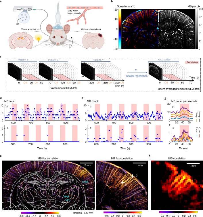
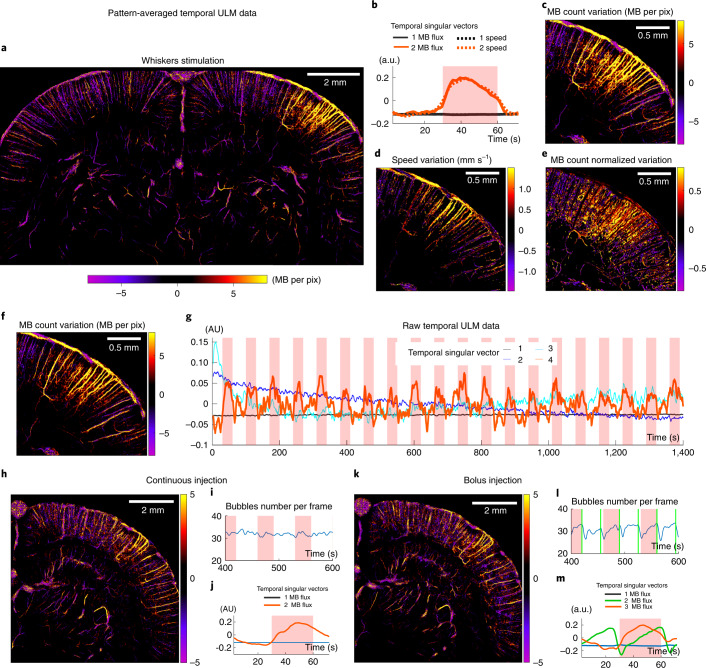
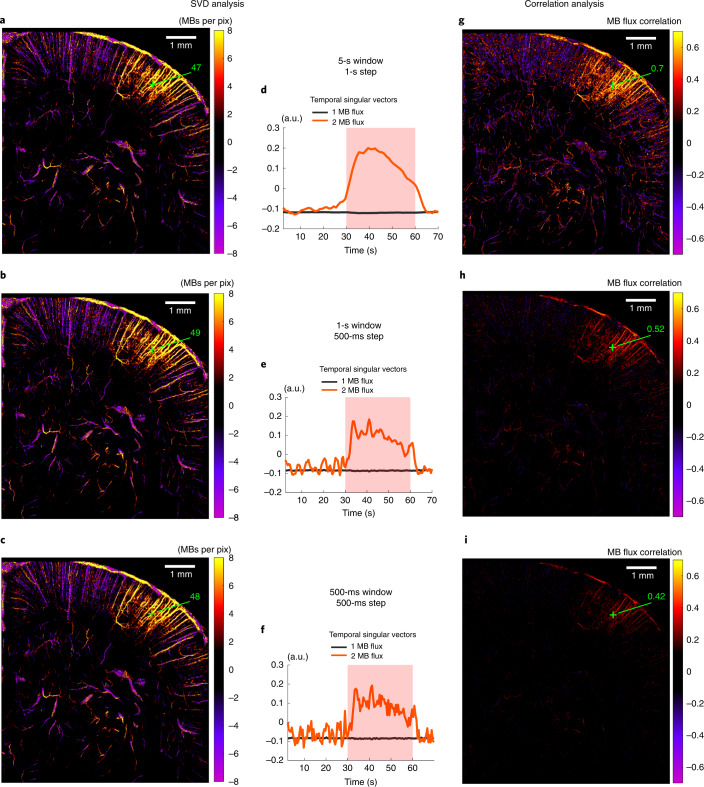
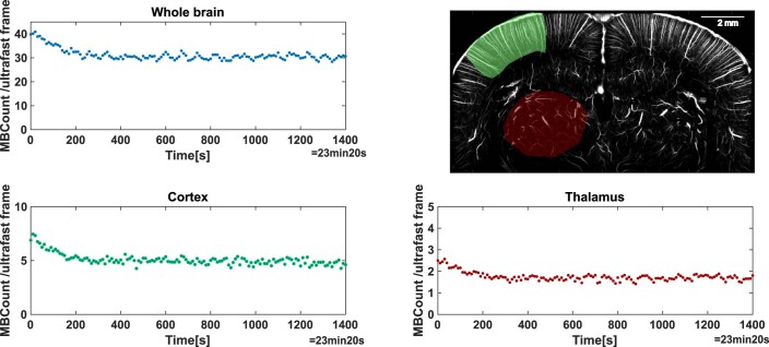
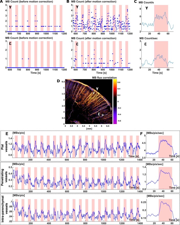
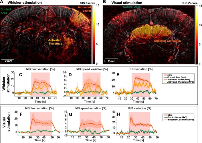
The advent of neuroimaging has increased our understanding of brain function. While most brain-wide functional imaging modalities exploit neurovascular coupling to map brain activity at millimeter resolutions, the recording of functional responses at microscopic scale in mammals remains the privilege of invasive electrophysiological or optical approaches, but is mostly restricted to either the cortical surface or the vicinity of implanted sensors. Ultrasound localization microscopy (ULM) has achieved transcranial imaging of cerebrovascular flow, up to micrometre scales, by localizing intravenously injected microbubbles; however, the long acquisition time required to detect microbubbles within microscopic vessels has so far restricted ULM application mainly to microvasculature structural imaging. Here we show how ULM can be modified to quantify functional hyperemia dynamically during brain activation reaching a 6.5-µm spatial and 1-s temporal resolution in deep regions of the rat brain.
神经影像学的出现增加了我们对大脑功能的理解。虽然大多数全脑功能成像模式都利用神经血管耦合来以毫米级分辨率绘制大脑活动图,但在哺乳动物中以微观尺度记录功能反应仍然是侵入性电生理或光学方法的特权,但主要限于皮质表面或植入传感器附近。超声定位显微镜(ULM)通过定位静脉内注射的微泡,实现了可达微米级的脑血管流的颅穿透成像;然而,由于在微观血管内检测微泡所需的长时间采集,迄今为止,ULM 的应用主要限于微血管结构成像。在这里,我们展示了如何修改 ULM 以在大脑激活期间动态量化功能充血,在大鼠大脑的深部区域达到 6.5-µm 的空间和 1-s 的时间分辨率。