• 文献检索
  • 文档翻译
  • 深度研究
  • 学术资讯
  • Suppr Zotero 插件Zotero 插件
  • 邀请有礼
  • 套餐&价格
  • 历史记录
应用&插件
Suppr Zotero 插件Zotero 插件浏览器插件Mac 客户端Windows 客户端微信小程序
定价
高级版会员购买积分包购买API积分包
服务
文献检索文档翻译深度研究API 文档MCP 服务
关于我们
关于 Suppr公司介绍联系我们用户协议隐私条款
关注我们

Suppr 超能文献

核心技术专利:CN118964589B侵权必究
粤ICP备2023148730 号-1Suppr @ 2026

文献检索

告别复杂PubMed语法,用中文像聊天一样搜索,搜遍4000万医学文献。AI智能推荐,让科研检索更轻松。

立即免费搜索

文件翻译

保留排版,准确专业,支持PDF/Word/PPT等文件格式,支持 12+语言互译。

免费翻译文档

深度研究

AI帮你快速写综述,25分钟生成高质量综述,智能提取关键信息,辅助科研写作。

立即免费体验

基于术前双层探测器光谱计算机断层扫描基于3D VOI的定量参数预测胰腺导管腺癌高Ki-67增殖指数模型的开发与验证

Development and validation of a model based on preoperative dual-layer detector spectral computed tomography 3D VOI-based quantitative parameters to predict high Ki-67 proliferation index in pancreatic ductal adenocarcinoma.

作者信息

Zeng Dan, Zhang Jiayan, Song Zuhua, Li Qian, Zhang Dan, Li Xiaojiao, Wen Youjia, Ren Xiaofang, Wang Xinwei, Zhang Xiaodi, Tang Zhuoyue

机构信息

Department of Radiology, Chongqing General Hospital, Chongqing University, Chongqing, China.

Department of Clinical and Technical Support, Philips Healthcare, Chengdu, China.

出版信息

Insights Imaging. 2024 Dec 5;15(1):291. doi: 10.1186/s13244-024-01864-9.

DOI:10.1186/s13244-024-01864-9
PMID:39636501
原文链接:https://pmc.ncbi.nlm.nih.gov/articles/PMC11621245/
Abstract

OBJECTIVE

To develop and validate a model integrating dual-layer detector spectral computed tomography (DLCT) three-dimensional (3D) volume of interest (VOI)-based quantitative parameters and clinical features for predicting Ki-67 proliferation index (PI) in pancreatic ductal adenocarcinoma (PDAC).

MATERIALS AND METHODS

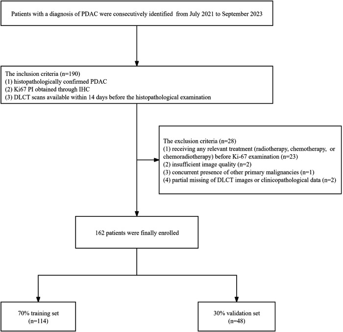
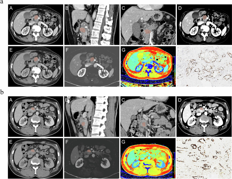
A total of 162 patients with histopathologically confirmed PDAC who underwent DLCT examination were included and allocated to the training (114) and validation (48) sets. 3D VOI-iodine concentration (IC), 3D VOI-slope of the spectral attenuation curves, and 3D VOI-effective atomic number were obtained from the portal venous phase. The significant clinical features and DLCT quantitative parameters were identified through univariate analysis and multivariate logistic regression. The discrimination capability and clinical applicability of the clinical, DLCT, and DLCT-clinical models were quantified by the Receiver Operating Characteristic curve (ROC) and Decision Curve Analysis (DCA), respectively. The optimal model was then used to develop a nomogram, with the goodness-of-fit evaluated through the calibration curve.

RESULTS

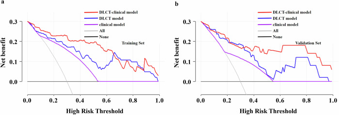
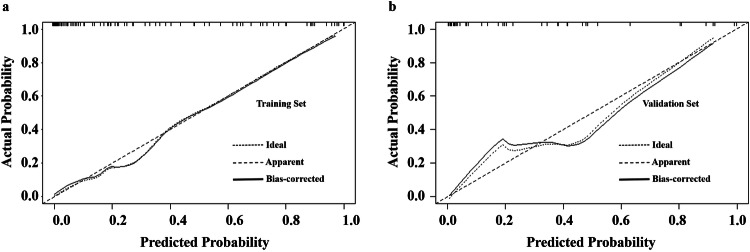
The DLCT-clinical model demonstrated superior predictive capability and a satisfactory net benefit for Ki-67 PI in PDAC compared to the clinical and DLCT models. The DLCT-clinical model integrating 3D VOI-IC and CA125 showed area under the ROC curves of 0.939 (95% CI, 0.895-0.982) and 0.915 (95% CI, 0.834-0.996) in the training and validation sets, respectively. The nomogram derived from the DLCT-clinical model exhibited favorable calibration, as depicted by the calibration curve.

CONCLUSIONS

The proposed model based on DLCT 3D VOI-IC and CA125 is a non-invasive and effective preoperative prediction tool demonstrating favorable predictive performance for Ki-67 PI in PDAC.

CRITICAL RELEVANCE STATEMENT

The dual-layer detector spectral computed tomography-clinical model could help predict high Ki-67 PI in pancreatic ductal adenocarcinoma patients, which may help clinicians provide appropriate and individualized treatments.

KEY POINTS

Dual-layer detector spectral CT (DLCT) could predict Ki-67 in pancreatic ductal adenocarcinoma (PDAC). The DLCT-clinical model improved the differential diagnosis of Ki-67. The nomogram showed satisfactory calibration and net benefit for discriminating Ki-67.

摘要

目的

建立并验证一种整合双层探测器光谱计算机断层扫描(DLCT)基于三维(3D)感兴趣区(VOI)的定量参数和临床特征的模型,用于预测胰腺导管腺癌(PDAC)中的Ki-67增殖指数(PI)。

材料与方法

纳入162例经组织病理学确诊且接受DLCT检查的PDAC患者,并将其分为训练集(114例)和验证集(48例)。在门静脉期获取3D VOI碘浓度(IC)、3D VOI光谱衰减曲线斜率和3D VOI有效原子序数。通过单因素分析和多因素逻辑回归确定显著的临床特征和DLCT定量参数。分别通过受试者操作特征曲线(ROC)和决策曲线分析(DCA)对临床、DLCT和DLCT-临床模型的鉴别能力和临床适用性进行量化。然后使用最优模型制定列线图,并通过校准曲线评估拟合优度。

结果

与临床和DLCT模型相比,DLCT-临床模型在预测PDAC中Ki-67 PI方面表现出卓越的预测能力和令人满意的净效益。整合3D VOI-IC和CA125的DLCT-临床模型在训练集和验证集中的ROC曲线下面积分别为0.939(95%CI,0.895-0.982)和0.915(95%CI,0.834-0.996)。如校准曲线所示,由DLCT-临床模型得出的列线图显示出良好的校准。

结论

所提出的基于DLCT 3D VOI-IC和CA125的模型是一种无创且有效的术前预测工具,对PDAC中的Ki-67 PI具有良好的预测性能。

关键相关性声明

双层探测器光谱计算机断层扫描-临床模型有助于预测胰腺导管腺癌患者的高Ki-67 PI,这可能有助于临床医生提供适当的个体化治疗。

关键点

双层探测器光谱CT(DLCT)可预测胰腺导管腺癌(PDAC)中的Ki-67。DLCT-临床模型改善了对Ki-67的鉴别诊断。列线图在鉴别Ki-67方面显示出令人满意的校准和净效益。

https://cdn.ncbi.nlm.nih.gov/pmc/blobs/720c/11621245/8ccde819f5aa/13244_2024_1864_Fig6_HTML.jpg
https://cdn.ncbi.nlm.nih.gov/pmc/blobs/720c/11621245/f9aed6993e45/13244_2024_1864_Fig1_HTML.jpg
https://cdn.ncbi.nlm.nih.gov/pmc/blobs/720c/11621245/5f84b809f668/13244_2024_1864_Fig2_HTML.jpg
https://cdn.ncbi.nlm.nih.gov/pmc/blobs/720c/11621245/ec0f22b94724/13244_2024_1864_Fig3_HTML.jpg
https://cdn.ncbi.nlm.nih.gov/pmc/blobs/720c/11621245/0eb262625563/13244_2024_1864_Fig4_HTML.jpg
https://cdn.ncbi.nlm.nih.gov/pmc/blobs/720c/11621245/a69255c017e3/13244_2024_1864_Fig5_HTML.jpg
https://cdn.ncbi.nlm.nih.gov/pmc/blobs/720c/11621245/8ccde819f5aa/13244_2024_1864_Fig6_HTML.jpg

相似文献

1
Development and validation of a model based on preoperative dual-layer detector spectral computed tomography 3D VOI-based quantitative parameters to predict high Ki-67 proliferation index in pancreatic ductal adenocarcinoma.基于术前双层探测器光谱计算机断层扫描基于3D VOI的定量参数预测胰腺导管腺癌高Ki-67增殖指数模型的开发与验证
Insights Imaging. 2024 Dec 5;15(1):291. doi: 10.1186/s13244-024-01864-9.
2
Radiomics analysis of dual-layer detector spectral CT-derived iodine maps for predicting Ki-67 PI in pancreatic ductal adenocarcinoma.双层探测器光谱CT衍生碘图用于预测胰腺导管腺癌中Ki-67增殖指数的影像组学分析
BMC Med Imaging. 2025 Apr 17;25(1):124. doi: 10.1186/s12880-025-01664-7.
3
A nomogram based on dual-layer detector spectral computed tomography quantitative parameters and morphological quantitative indicator for distinguishing metastatic and nonmetastatic regional lymph nodes in pancreatic ductal adenocarcinoma.基于双层探测器光谱计算机断层扫描定量参数和形态学定量指标的列线图,用于鉴别胰腺导管腺癌区域淋巴结转移与非转移情况。
Quant Imaging Med Surg. 2024 Jul 1;14(7):4376-4387. doi: 10.21037/qims-23-1624. Epub 2024 May 31.
4
The preoperative prediction of lymph node metastasis of resectable pancreatic ductal adenocarcinoma using dual-layer spectral computed tomography.使用双层光谱计算机断层扫描对可切除性胰腺导管腺癌的淋巴结转移进行术前预测。
Eur Radiol. 2025 May;35(5):2692-2701. doi: 10.1007/s00330-024-11143-2. Epub 2024 Oct 24.
5
Development and validation of a model for predicting the expression of Ki-67 in pancreatic ductal adenocarcinoma with radiological features and dual-energy computed tomography quantitative parameters.利用放射学特征和双能计算机断层扫描定量参数预测胰腺导管腺癌中Ki-67表达的模型的开发与验证
Insights Imaging. 2024 Feb 14;15(1):41. doi: 10.1186/s13244-024-01617-8.
6
A clinical-radiomics nomogram based on dual-layer spectral detector CT to predict cancer stage in pancreatic ductal adenocarcinoma.基于双层光谱探测器 CT 的临床放射组学列线图预测胰腺导管腺癌的癌症分期。
Cancer Imaging. 2024 May 9;24(1):55. doi: 10.1186/s40644-024-00700-z.
7
Development of a CT radiomics nomogram for preoperative prediction of Ki-67 index in pancreatic ductal adenocarcinoma: a two-center retrospective study.基于 CT 影像组学构建用于预测胰腺导管腺癌 Ki-67 指数的列线图:一项多中心回顾性研究。
Eur Radiol. 2024 May;34(5):2934-2943. doi: 10.1007/s00330-023-10393-w. Epub 2023 Nov 8.
8
Detection of malignant lesions in cytologically indeterminate thyroid nodules using a dual-layer spectral detector CT-clinical nomogram.使用双层光谱探测器CT临床列线图检测甲状腺细针穿刺结果不确定的结节中的恶性病变
Front Oncol. 2024 May 28;14:1357419. doi: 10.3389/fonc.2024.1357419. eCollection 2024.
9
A prediction model based on dual-layer spectral detector computed tomography for distinguishing nonluminal from luminal invasive breast cancer.基于双层光谱探测器计算机断层扫描的鉴别非腔内与腔内浸润性乳腺癌的预测模型。
Quant Imaging Med Surg. 2024 Dec 5;14(12):8672-8685. doi: 10.21037/qims-24-598. Epub 2024 Nov 11.
10
Dual-layer spectral detector CT: A noninvasive preoperative tool for predicting histopathological differentiation in pancreatic ductal adenocarcinoma.双层光谱探测器CT:一种用于预测胰腺导管腺癌组织病理学分化的无创术前工具。
Eur J Radiol. 2024 Apr;173:111327. doi: 10.1016/j.ejrad.2024.111327. Epub 2024 Jan 24.

引用本文的文献

1
Radiomics analysis of dual-layer detector spectral CT-derived iodine maps for predicting Ki-67 PI in pancreatic ductal adenocarcinoma.双层探测器光谱CT衍生碘图用于预测胰腺导管腺癌中Ki-67增殖指数的影像组学分析
BMC Med Imaging. 2025 Apr 17;25(1):124. doi: 10.1186/s12880-025-01664-7.

本文引用的文献

1
Prognostic significance of grade of malignancy based on histopathological differentiation and Ki-67 in pancreatic ductal adenocarcinoma.基于组织病理学分化和 Ki-67 评估胰腺导管腺癌恶性程度分级的预后意义。
Cancer Biol Med. 2024 Jan 3;21(5):416-32. doi: 10.20892/j.issn.2095-3941.2023.0363.
2
Combined utility of Ki-67 index and tumor grade to stratify patients with pancreatic ductal adenocarcinoma who underwent upfront surgery.Ki-67 指数与肿瘤分级联合用于分层接受直接手术的胰腺导管腺癌患者。
BMC Surg. 2023 Dec 8;23(1):370. doi: 10.1186/s12893-023-02256-4.
3
Correlation between Spectral CT Parameters and Ki67 Expression in Hepatocellular Carcinoma.
肝细胞癌光谱CT参数与Ki67表达的相关性
Curr Med Imaging. 2023 Oct 14. doi: 10.2174/0115734056227641231004042735.
4
Usefulness of Three-Dimensional Iodine Mapping Quantified by Dual-Energy CT for Differentiating Thymic Epithelial Tumors.双能CT定量三维碘映射在鉴别胸腺上皮肿瘤中的应用价值
J Clin Med. 2023 Aug 28;12(17):5610. doi: 10.3390/jcm12175610.
5
Quantitative parameters in novel spectral computed tomography: Assessment of Ki-67 expression in patients with gastric adenocarcinoma.新型光谱 CT 中的定量参数:胃腺癌患者 Ki-67 表达的评估。
World J Gastroenterol. 2023 Mar 14;29(10):1602-1613. doi: 10.3748/wjg.v29.i10.1602.
6
Differentiating between pulmonary adenocarcinoma and squamous cell carcinoma by spectral CT volumetric quantitative analysis: a comparative study with conventional spectral analysis.通过光谱CT容积定量分析鉴别肺腺癌和鳞状细胞癌:与传统光谱分析的对比研究
J Thorac Dis. 2023 Feb 28;15(2):679-689. doi: 10.21037/jtd-23-115. Epub 2023 Feb 22.
7
Reproducibility of 2D versus 3D radiomics for quantitative assessment of fetal lung development: a retrospective fetal MRI study.二维与三维影像组学在胎儿肺发育定量评估中的可重复性:一项回顾性胎儿磁共振成像研究
Insights Imaging. 2023 Feb 8;14(1):31. doi: 10.1186/s13244-023-01376-y.
8
Cancer statistics, 2023.癌症统计数据,2023 年。
CA Cancer J Clin. 2023 Jan;73(1):17-48. doi: 10.3322/caac.21763.
9
Correlation Between Quantitative Spectral CT Parameters and Ki-67 Expression in Lung Adenocarcinomas Manifesting as Ground-glass Nodules.定量光谱 CT 参数与表现为磨玻璃结节的肺腺癌 Ki-67 表达的相关性。
Curr Med Imaging. 2023;19(9):1052-1062. doi: 10.2174/1573405619666221230120528.
10
Development and validation of a nomogram to predict liver metastasis for pancreatic ductal adenocarcinoma after radical resection.预测根治性切除术后胰腺导管腺癌肝转移的列线图的开发与验证
Front Oncol. 2022 Nov 21;12:1040411. doi: 10.3389/fonc.2022.1040411. eCollection 2022.