Zeng Dan, Song Zuhua, Liu Qian, Huang Jie, Wang Xinwei, Tang Zhuoyue
Department of Radiology, Chongqing General Hospital, Chongqing, China.
BMC Med Imaging. 2025 Apr 17;25(1):124. doi: 10.1186/s12880-025-01664-7.
To evaluate the feasibility of radiomics analysis using dual-layer detector spectral CT (DLCT)-derived iodine maps for the preoperative prediction of the Ki-67 proliferation index (PI) in pancreatic ductal adenocarcinoma (PDAC).
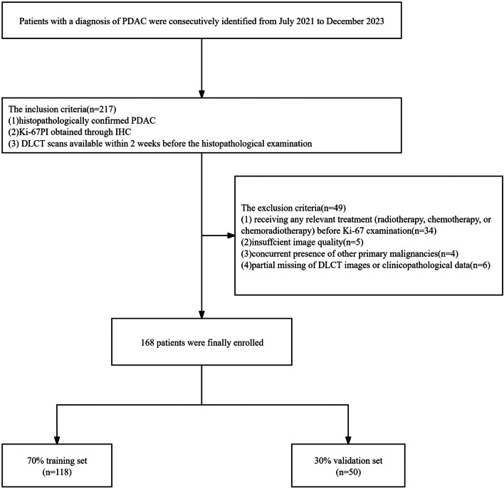
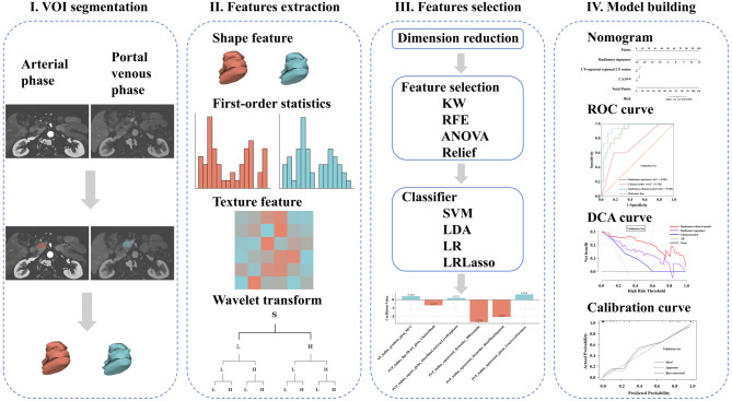
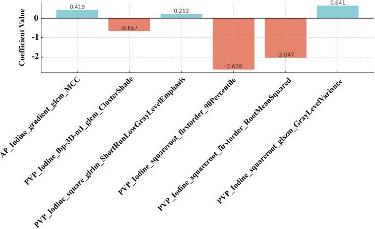
A total of 168 PDAC patients who underwent DLCT examination were included and randomly allocated to the training (n = 118) and validation (n = 50) sets. A clinical model was constructed using independent clinicoradiological features identified through multivariate logistic regression analysis in the training set. The radiomics signature was generated based on the coefficients of selected features from iodine maps in the arterial and portal venous phases of DLCT. Finally, a radiomics-clinical model was developed by integrating the radiomics signature and significant clinicoradiological features. The predictive performance of three models was evaluated using the Receiver Operating Characteristic (ROC) curve and Decision Curve Analysis. The optimal model was then used to develop a nomogram, with goodness-of-fit evaluated through the calibration curve.
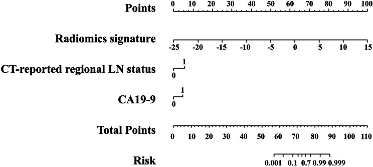
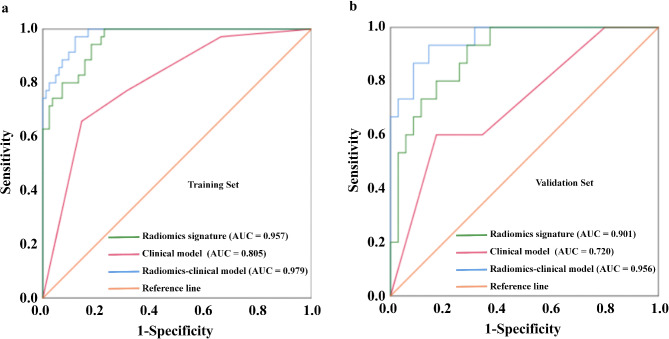
The radiomics-clinical model integrating radiomics signature, CA19-9, and CT-reported regional lymph node status demonstrated excellent performance in predicting Ki-67 PI in PDAC, which showed an area under the ROC curve of 0.979 and 0.956 in the training and validation sets, respectively. The radiomics-clinical nomogram demonstrated the improved net benefit and exhibited satisfactory consistency.
This exploratory study demonstrated the feasibility of using DLCT-derived iodine map-based radiomics to predict Ki-67 PI preoperatively in PDAC patients. While preliminary, our findings highlight the potential of functional imaging combined with radiomics for personalized treatment planning.
评估使用双层探测器光谱CT(DLCT)衍生的碘图进行影像组学分析以术前预测胰腺导管腺癌(PDAC)中Ki-67增殖指数(PI)的可行性。
纳入168例接受DLCT检查的PDAC患者,并随机分为训练组(n = 118)和验证组(n = 50)。在训练组中,利用多因素逻辑回归分析确定的独立临床放射学特征构建临床模型。基于DLCT动脉期和门静脉期碘图中选定特征的系数生成影像组学特征。最后,通过整合影像组学特征和显著的临床放射学特征建立影像组学-临床模型。使用受试者操作特征(ROC)曲线和决策曲线分析评估三种模型的预测性能。然后使用最佳模型制定列线图,并通过校准曲线评估拟合优度。
整合影像组学特征、CA19-9和CT报告的区域淋巴结状态的影像组学-临床模型在预测PDAC中Ki-67 PI方面表现出色,训练组和验证组的ROC曲线下面积分别为0.979和0.956。影像组学-临床列线图显示净效益提高且一致性良好。
这项探索性研究证明了使用基于DLCT衍生碘图的影像组学术前预测PDAC患者Ki-67 PI的可行性。虽然是初步研究,但我们的发现突出了功能成像结合影像组学在个性化治疗规划中的潜力。