Onagi Akifumi, Sugimoto Kotaro, Kobayashi Makoto, Sato Yumi, Kobayashi Yasuyuki, Yaginuma Kei, Meguro Satoru, Hoshi Seiji, Hata Jyunya, Hashimoto Yuko, Kojima Yoshiyuki, Chiba Hideki
Department of Basic Pathology, Fukushima Medical University School of Medicine, Fukushima, 960-1295, Japan.
Department of Urology, Fukushima Medical University School of Medicine, Fukushima, 960-1295, Japan.
Cell Commun Signal. 2024 Dec 5;22(1):588. doi: 10.1186/s12964-024-01964-5.
BACKGROUND & AIMS: In addition to their adhesive properties, cell adhesion molecules such as claudins (CLDNs) exhibit signaling ability to organize diverse cellular events. Although the CLDN-adhesion signaling stimulates or inhibits cancer progression, the underlying mechanism remains poorly established. Here, we verified whether and how CLDN10 promotes intracellular signals and malignant phenotypes in clear cell renal cell carcinoma (ccRCC).
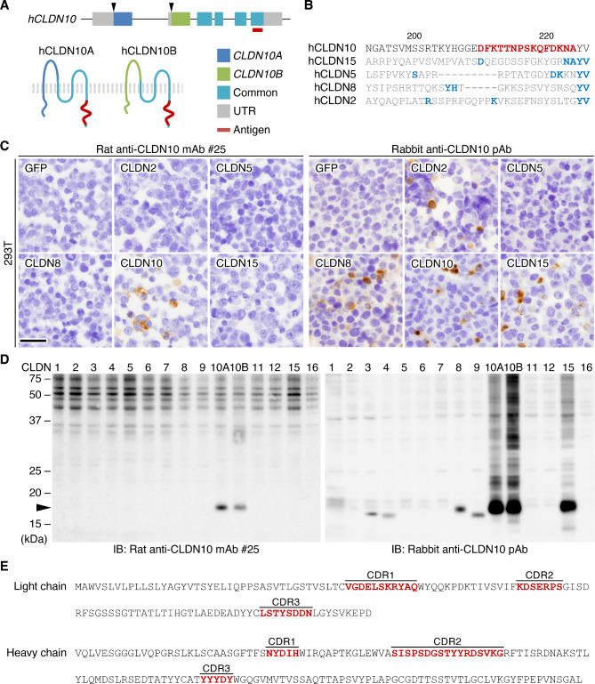
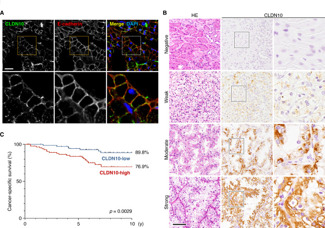
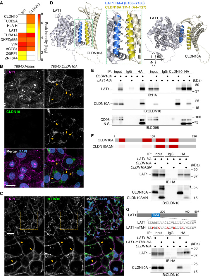
We developed a novel monoclonal antibody that specifically recognizes CLDN10. By immunohistochemistry using this antibody, the clinicopathological significance of aberrant CLDN10 expression in 165 ccRCC patients was determined. We next generated the ccRCC cells (786-O, ACHN, and OS-RC-2) expressing CLDN10, and compared their phenotypes with those of control cells. Immunoprecipitation-mass spectrometry was used to identify a CLDN10-interacting protein, followed by evaluation of its association with CLDN10 and loss-of-functions in ccRCC cells.
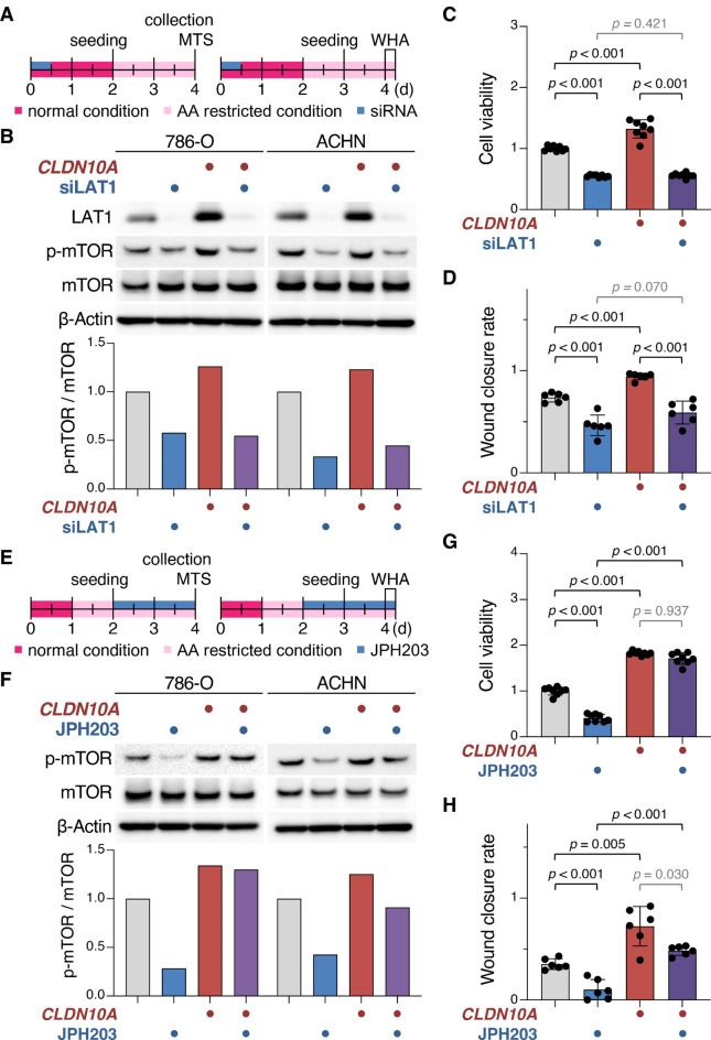
High CLDN10 expression predicted poor outcome in ccRCC patients and represented an independent prognostic marker for cancer-specific survival. Cell surface CLDN10 promoted cell viability, proliferation, and migration of ccRCC cells, as well as their tumor growth. CLDN10 also activated mTOR signaling and expression of downstream targets, including MYC target genes. Notably, we found that CLDN10 forms a complex with an amino acid transporter, LAT1, and that CLDN10-LAT1 signaling facilitates malignant phenotypes in ccRCC cells. Structural prediction and immunoprecipitation analysis results strongly suggest an interaction between CLDN10-TM1 (transmembrane domain 1) and LAT1-TM4.
We conclude that CLDN10-LAT1 signaling drives ccRCC progression. Taken together with our previous findings on CLDN-Src-family kinases signaling, CLDNs propagate distinct intracellular signals depending on their association with different binding partners.
除了具有黏附特性外,紧密连接蛋白(CLDNs)等细胞黏附分子还具有组织多种细胞活动的信号传导能力。尽管CLDN黏附信号传导可刺激或抑制癌症进展,但其潜在机制仍不清楚。在此,我们验证了CLDN10是否以及如何促进透明细胞肾细胞癌(ccRCC)中的细胞内信号传导和恶性表型。
我们开发了一种特异性识别CLDN10的新型单克隆抗体。通过使用该抗体进行免疫组织化学,确定了165例ccRCC患者中异常CLDN10表达的临床病理意义。接下来,我们构建了表达CLDN10的ccRCC细胞(786-O、ACHN和OS-RC-2),并将它们的表型与对照细胞进行比较。采用免疫沉淀-质谱法鉴定与CLDN10相互作用的蛋白,随后评估其与CLDN10的关联以及在ccRCC细胞中的功能丧失情况。
CLDN10高表达预示ccRCC患者预后不良,是癌症特异性生存的独立预后标志物。细胞表面的CLDN10促进ccRCC细胞的活力、增殖、迁移及其肿瘤生长。CLDN10还激活mTOR信号传导和下游靶点的表达,包括MYC靶基因。值得注意的是,我们发现CLDN10与一种氨基酸转运体LAT1形成复合物,并且CLDN10-LAT1信号传导促进ccRCC细胞中的恶性表型。结构预测和免疫沉淀分析结果强烈表明CLDN10跨膜结构域1(TM1)与LAT1跨膜结构域4(TM4)之间存在相互作用。
我们得出结论,CLDN10-LAT1信号传导驱动ccRCC进展。结合我们之前关于CLDN- src家族激酶信号传导的研究结果,CLDNs根据其与不同结合伙伴的关联传递不同的细胞内信号。