• 文献检索
  • 文档翻译
  • 深度研究
  • 学术资讯
  • Suppr Zotero 插件Zotero 插件
  • 邀请有礼
  • 套餐&价格
  • 历史记录
应用&插件
Suppr Zotero 插件Zotero 插件浏览器插件Mac 客户端Windows 客户端微信小程序
定价
高级版会员购买积分包购买API积分包
服务
文献检索文档翻译深度研究API 文档MCP 服务
关于我们
关于 Suppr公司介绍联系我们用户协议隐私条款
关注我们

Suppr 超能文献

核心技术专利:CN118964589B侵权必究
粤ICP备2023148730 号-1Suppr @ 2026

文献检索

告别复杂PubMed语法,用中文像聊天一样搜索,搜遍4000万医学文献。AI智能推荐,让科研检索更轻松。

立即免费搜索

文件翻译

保留排版,准确专业,支持PDF/Word/PPT等文件格式,支持 12+语言互译。

免费翻译文档

深度研究

AI帮你快速写综述,25分钟生成高质量综述,智能提取关键信息,辅助科研写作。

立即免费体验

强直性肌营养不良患者心脏磁共振成像评估心脏风险及心肌纤维化

Cardiac risk and myocardial fibrosis assessment with cardiac magnetic resonance in patients with myotonic dystrophy.

作者信息

Abati Elena, Alberti Claudia, Tambè Valentina, Esseridou Anastasia, Comi Giacomo Pietro, Corti Stefania, Meola Giovanni, Secchi Francesco

机构信息

Department of Pathophysiology and Transplantation, Dino Ferrari Center, Università degli Studi di Milano, Milan, Italy.

Neurology Unit, Foundation IRCCS Ca' Granda Ospedale Maggiore Policlinico, Milan, Italy.

出版信息

Front Neurol. 2024 Nov 21;15:1493570. doi: 10.3389/fneur.2024.1493570. eCollection 2024.

DOI:10.3389/fneur.2024.1493570
PMID:39639989
原文链接:https://pmc.ncbi.nlm.nih.gov/articles/PMC11617365/
Abstract

INTRODUCTION

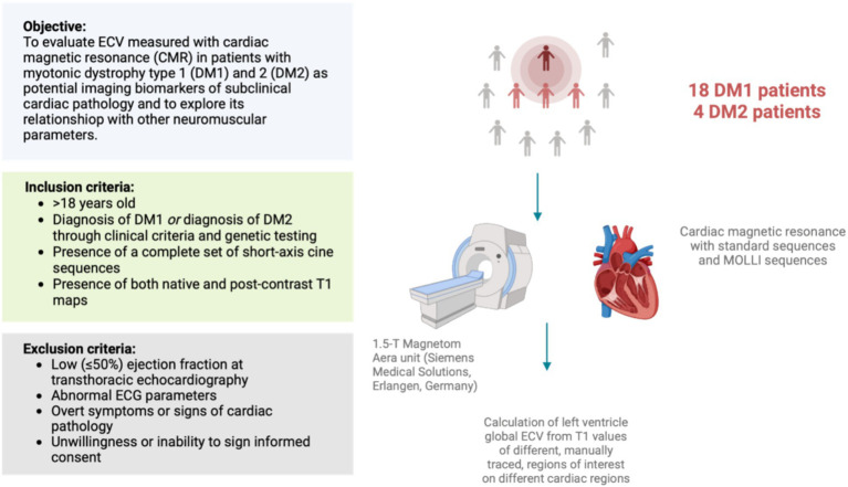
Non-invasive evaluation of myocardial tissue is a major goal of cardiac imaging. This is the case of myocardial fibrosis which is crucial in many myocardial diseases. Cardiac extracellular volume (ECV) was shown to indicate myocardial fibrosis and early cardiac involvement. With this study, our objective is to evaluate ECV measured with cardiac magnetic resonance (CMR) in patients with myotonic dystrophy type 1 (DM1) and 2 (DM2) as potential imaging biomarkers of subclinical cardiac pathology, and its relationship with demographic and clinical parameters, ECG-derived measures of cardiac conduction, and neuromuscular performance status.

MATERIALS AND METHODS

We retrospectively analyzed 18 DM1 patients and 4 DM2 patients without apparent cardiac disease who had CMR at our center. Differences between independent distributions were evaluated using Mann-Whitney U test, while correlations were evaluated using Spearman's .

RESULTS

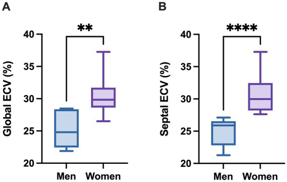
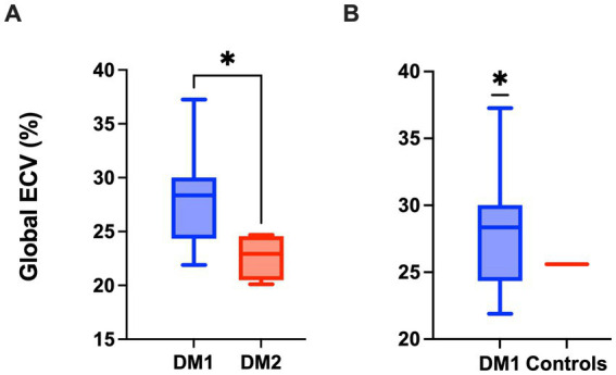
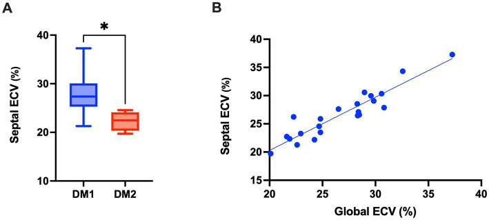
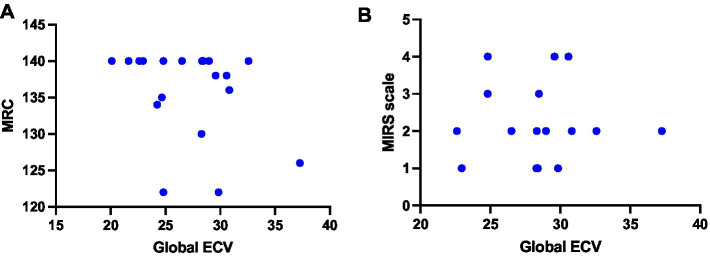
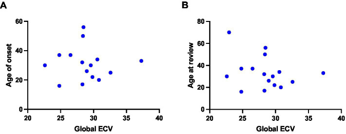
Global ECV in DM1 patients (median 28.36; IQR 24.81-29.77) was significantly higher ( = 0.0141) than in DM2 patients (median 22.93; IQR 21.25-24.35), and than that reported in literature in healthy subjects ( = 0.0374; median 25.60; IQR 19.90-31.90). Septal ECV was significantly higher ( = 0.0074) in DM1 (median 27.37; IQR 25.97-29.74) than in DM2 patients (median 22.46; 21.57-23.19). Global ECV showed a strong, positive correlation with septal ECV ( = 0.9282,  < 0.0001). We observed that DM1 women showed significantly higher global ( = 0.0012) and septal ( < 0.0001) ECV values compared to men.

DISCUSSION

We found a significant increase in global and septal cardiac ECV in patients with DM1. These values might thus suggest that DM1 patients present an increased cardiovascular risk, mainly due to cardiac fibrosis, even in absence of overt cardiac pathology at other common cardiovascular exams. DM1 patients may also be at increased risk of early septal fibrosis, with important implications on the risk for fatal arrhythmias. In addition, our results suggest the presence of gender-related differences, with DM1 women being more prone to myocardial fibrosis. Physicians dealing with DM1 may consider CMR as a screening tool for the early identification of patients with increased cardiovascular risk.

摘要

引言

心肌组织的无创评估是心脏成像的主要目标。在许多心肌疾病中至关重要的心肌纤维化就是这种情况。心脏细胞外容积(ECV)已被证明可指示心肌纤维化和早期心脏受累情况。通过本研究,我们的目的是评估在1型强直性肌营养不良(DM1)和2型强直性肌营养不良(DM2)患者中利用心脏磁共振成像(CMR)测量的ECV,将其作为亚临床心脏病理的潜在成像生物标志物,并评估其与人口统计学和临床参数、心电图衍生的心脏传导测量值以及神经肌肉功能状态之间的关系。

材料与方法

我们回顾性分析了在我们中心接受CMR检查的18例无明显心脏疾病的DM1患者和4例DM2患者。使用曼-惠特尼U检验评估独立分布之间的差异,同时使用斯皮尔曼相关系数评估相关性。

结果

DM1患者的整体ECV(中位数28.36;四分位数间距24.81 - 29.77)显著高于DM2患者(中位数22.93;四分位数间距21.25 - 24.35)(P = 0.0141),也高于文献报道的健康受试者的ECV(P = 0.0374;中位数25.60;四分位数间距19.90 - 31.90)。DM1患者的室间隔ECV(中位数27.37;四分位数间距25.97 - 29.74)显著高于DM2患者(中位数22.46;21.57 - 23.19)(P = 0.0074)。整体ECV与室间隔ECV呈强正相关(r = 0.9282,P < 0.0001)。我们观察到,与男性相比,DM1女性的整体(P = 0.0012)和室间隔(P < 0.0001)ECV值显著更高。

讨论

我们发现DM1患者的整体和室间隔心脏ECV显著增加。因此,这些值可能表明DM1患者存在心血管风险增加的情况,主要是由于心脏纤维化,即使在其他常见心血管检查中没有明显心脏病理表现。DM1患者早期室间隔纤维化的风险也可能增加,这对致命性心律失常的风险有重要影响。此外,我们的结果表明存在性别相关差异,DM1女性更容易发生心肌纤维化。处理DM1的医生可将CMR视为早期识别心血管风险增加患者的筛查工具。

https://cdn.ncbi.nlm.nih.gov/pmc/blobs/e028/11617365/f457fed258ea/fneur-15-1493570-g006.jpg
https://cdn.ncbi.nlm.nih.gov/pmc/blobs/e028/11617365/00175568198b/fneur-15-1493570-g001.jpg
https://cdn.ncbi.nlm.nih.gov/pmc/blobs/e028/11617365/348b47538f9f/fneur-15-1493570-g002.jpg
https://cdn.ncbi.nlm.nih.gov/pmc/blobs/e028/11617365/0bf1294a6937/fneur-15-1493570-g003.jpg
https://cdn.ncbi.nlm.nih.gov/pmc/blobs/e028/11617365/6aeb7cd08143/fneur-15-1493570-g004.jpg
https://cdn.ncbi.nlm.nih.gov/pmc/blobs/e028/11617365/f5477555b823/fneur-15-1493570-g005.jpg
https://cdn.ncbi.nlm.nih.gov/pmc/blobs/e028/11617365/f457fed258ea/fneur-15-1493570-g006.jpg

相似文献

1
Cardiac risk and myocardial fibrosis assessment with cardiac magnetic resonance in patients with myotonic dystrophy.强直性肌营养不良患者心脏磁共振成像评估心脏风险及心肌纤维化
Front Neurol. 2024 Nov 21;15:1493570. doi: 10.3389/fneur.2024.1493570. eCollection 2024.
2
Rare Disease: Cardiac Risk Assessment With MRI in Patients With Myotonic Dystrophy Type 1.罕见病:1型强直性肌营养不良患者心脏风险的磁共振成像评估
Front Neurol. 2020 Mar 19;11:192. doi: 10.3389/fneur.2020.00192. eCollection 2020.
3
Myocardial fibrosis in patients with myotonic dystrophy type 1: a cardiovascular magnetic resonance study.1型强直性肌营养不良患者的心肌纤维化:一项心血管磁共振研究。
J Cardiovasc Magn Reson. 2014 Aug 1;16(1):59. doi: 10.1186/s12968-014-0059-z.
4
Myocardial fibrosis imaging based on T1-mapping and extracellular volume fraction (ECV) measurement in muscular dystrophy patients: diagnostic value compared with conventional late gadolinium enhancement (LGE) imaging.基于T1映射和细胞外容积分数(ECV)测量的肌营养不良患者心肌纤维化成像:与传统钆延迟增强(LGE)成像相比的诊断价值
Eur Heart J Cardiovasc Imaging. 2014 Sep;15(9):1004-12. doi: 10.1093/ehjci/jeu050. Epub 2014 Mar 30.
5
Progressive myocardial injury in myotonic dystrophy type II and facioscapulohumeral muscular dystrophy 1: a cardiovascular magnetic resonance follow-up study.进行性心肌损伤在肌强直性营养不良 2 型和面肩肱型肌营养不良 1 型中的表现:一项心血管磁共振随访研究。
J Cardiovasc Magn Reson. 2021 Nov 8;23(1):130. doi: 10.1186/s12968-021-00812-6.
6
Myocardial extracellular volume quantification in cardiac amyloidosis: a comparative study between cardiac computed tomography and magnetic resonance imaging.心肌细胞外容积在心脏淀粉样变性中的定量评估:心脏 CT 与 MRI 的对比研究。
Eur Radiol. 2024 Feb;34(2):1016-1025. doi: 10.1007/s00330-023-10129-w. Epub 2023 Aug 19.
7
Elevated myocardial extracellular volume fraction is associated with the development of conduction pathway defects following transcatheter aortic valve replacement.经导管主动脉瓣置换术后心肌细胞外体积分数升高与传导通路缺陷的发展有关。
Catheter Cardiovasc Interv. 2024 Nov;104(5):1119-1128. doi: 10.1002/ccd.31136. Epub 2024 Jul 2.
8
T-Mapping and extracellular volume estimates in pediatric subjects with Duchenne muscular dystrophy and healthy controls at 3T.3T 下杜氏肌营养不良症患儿与健康对照者的 T 映射和细胞外容积评估。
J Cardiovasc Magn Reson. 2020 Dec 10;22(1):85. doi: 10.1186/s12968-020-00687-z.
9
Myocardial fibrosis by late gadolinium enhancement cardiovascular magnetic resonance in myotonic muscular dystrophy type 1: highly prevalent but not associated with surface conduction abnormality.1 型强直性肌营养不良症的心脏磁共振钆延迟增强:心肌纤维化高度流行,但与体表传导异常无关。
J Cardiovasc Magn Reson. 2019 May 2;21(1):26. doi: 10.1186/s12968-019-0535-6.
10
Cardiac Pathology in Myotonic Dystrophy Type 1.1 型肌强直性营养不良的心脏病理学。
Int J Mol Sci. 2021 Nov 2;22(21):11874. doi: 10.3390/ijms222111874.

本文引用的文献

1
Sex-based differences in cardiovascular proteomic profiles and their associations with adverse outcomes in patients with chronic heart failure.基于性别的心血管蛋白质组谱差异及其与慢性心力衰竭患者不良结局的关系。
Biol Sex Differ. 2023 May 17;14(1):29. doi: 10.1186/s13293-023-00516-9.
2
Cardiac magnetic resonance findings and prognosis in type 1 myotonic dystrophy.1 型肌强直性营养不良的心脏磁共振表现与预后。
J Cardiovasc Med (Hagerstown). 2023 Jun 1;24(6):340-347. doi: 10.2459/JCM.0000000000001476. Epub 2023 Apr 27.
3
T1 values and extracellular volume fraction in asymptomatic subjects: variations in left ventricular segments and correlation with cardiovascular risk factors.
无症状受试者的 T1 值和细胞外容积分数:左心室节段的变化与心血管危险因素的相关性。
Sci Rep. 2022 Jul 22;12(1):12544. doi: 10.1038/s41598-022-16696-0.
4
Global Prevalence of Myotonic Dystrophy: An Updated Systematic Review and Meta-Analysis.全球强直性肌营养不良症患病率:一项更新的系统评价和荟萃分析。
Neuroepidemiology. 2022;56(3):163-173. doi: 10.1159/000524734. Epub 2022 Apr 28.
5
Cardiac Pathology in Myotonic Dystrophy Type 1.1 型肌强直性营养不良的心脏病理学。
Int J Mol Sci. 2021 Nov 2;22(21):11874. doi: 10.3390/ijms222111874.
6
Progressive myocardial injury in myotonic dystrophy type II and facioscapulohumeral muscular dystrophy 1: a cardiovascular magnetic resonance follow-up study.进行性心肌损伤在肌强直性营养不良 2 型和面肩肱型肌营养不良 1 型中的表现:一项心血管磁共振随访研究。
J Cardiovasc Magn Reson. 2021 Nov 8;23(1):130. doi: 10.1186/s12968-021-00812-6.
7
Myotonic dystrophy type 2: the 2020 update.肌强直性营养不良 2 型:2020 更新版。
Acta Myol. 2020 Dec 1;39(4):222-234. doi: 10.36185/2532-1900-026. eCollection 2020 Dec.
8
Rare Disease: Cardiac Risk Assessment With MRI in Patients With Myotonic Dystrophy Type 1.罕见病:1型强直性肌营养不良患者心脏风险的磁共振成像评估
Front Neurol. 2020 Mar 19;11:192. doi: 10.3389/fneur.2020.00192. eCollection 2020.
9
Clinical Care Recommendations for Cardiologists Treating Adults With Myotonic Dystrophy.临床护理建议:心内科医生治疗成年肌强直性营养不良患者
J Am Heart Assoc. 2020 Feb 18;9(4):e014006. doi: 10.1161/JAHA.119.014006. Epub 2020 Feb 6.
10
Consensus-based care recommendations for adults with myotonic dystrophy type 2.基于共识的2型强直性肌营养不良症成人护理建议。
Neurol Clin Pract. 2019 Aug;9(4):343-353. doi: 10.1212/CPJ.0000000000000645.