• 文献检索
  • 文档翻译
  • 深度研究
  • 学术资讯
  • Suppr Zotero 插件Zotero 插件
  • 邀请有礼
  • 套餐&价格
  • 历史记录
应用&插件
Suppr Zotero 插件Zotero 插件浏览器插件Mac 客户端Windows 客户端微信小程序
定价
高级版会员购买积分包购买API积分包
服务
文献检索文档翻译深度研究API 文档MCP 服务
关于我们
关于 Suppr公司介绍联系我们用户协议隐私条款
关注我们

Suppr 超能文献

核心技术专利:CN118964589B侵权必究
粤ICP备2023148730 号-1Suppr @ 2026

文献检索

告别复杂PubMed语法,用中文像聊天一样搜索,搜遍4000万医学文献。AI智能推荐,让科研检索更轻松。

立即免费搜索

文件翻译

保留排版,准确专业,支持PDF/Word/PPT等文件格式,支持 12+语言互译。

免费翻译文档

深度研究

AI帮你快速写综述,25分钟生成高质量综述,智能提取关键信息,辅助科研写作。

立即免费体验

罕见病:1型强直性肌营养不良患者心脏风险的磁共振成像评估

Rare Disease: Cardiac Risk Assessment With MRI in Patients With Myotonic Dystrophy Type 1.

作者信息

Alì Marco, Monti Caterina Beatrice, Melazzini Luca, Cardani Rosanna, Fossati Barbara, Cavalli Michele, Chow Kelvin, Secchi Francesco, Meola Giovanni, Sardanelli Francesco

机构信息

Unit of Diagnostic Imaging and Stereotactic Radiosurgery, C.D.I. Centro Diagnostico Italiano S.p.A., Milan, Italy.

Unit of Radiology, IRCCS Policlinico San Donato, San Donato Milanese, Italy.

出版信息

Front Neurol. 2020 Mar 19;11:192. doi: 10.3389/fneur.2020.00192. eCollection 2020.

DOI:10.3389/fneur.2020.00192
PMID:32265828
原文链接:https://pmc.ncbi.nlm.nih.gov/articles/PMC7098463/
Abstract

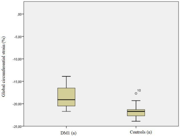
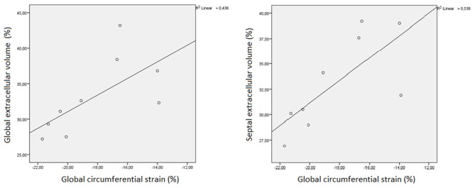
To evaluate myocardial strain and extracellular volume in myotonic dystrophy type 1 (DM1) patients as potential imaging biomarkers of subclinical cardiac pathology. We retrospectively analyzed 9 DM1 patients without apparent cardiac disease who had undergone cardiac magnetic resonance at our center. Patients were age- and sex-matched with healthy controls. The Mann-Whitney U test was used to compare cardiac strain between the two groups. The -test was used to compare the extracellular volume obtained in DM1 patients with that in healthy subject. Spearman's ρ was used for studying the associations among imaging parameters. Global cardiac strain (median -19.1%; IQR -20.5%, -16.5%) in DM1 patients was lower ( = 0.011) than that in controls (median-21.7%; IQR-22.7%,-21.3%). Global extracellular volume in DM1 patients (median 32.3%; IQR 29.3%,36.8%) was significantly ( = 0.008) higher than that reported in literature in healthy subjects (median 25.6%; IQR 19.9%,31.9%). Global cardiac strain showed a strong, positive correlation with septal strain (ρ = 0.767, = 0.016) and with both global (ρ = 0.733 = 0.025) and septal extracellular volume (ρ = 0.767, = 0.016). The increase in cardiac extracellular volume and decrease in strain are signs of early cardiac pathology in DM1. Physicians dealing with DM1 may take into consideration cardiac magnetic resonance as a screening tool to identify early cardiac involvement in this condition.

摘要

评估1型强直性肌营养不良(DM1)患者的心肌应变和细胞外容积,作为亚临床心脏病变的潜在影像学生物标志物。我们回顾性分析了9例在本中心接受心脏磁共振检查且无明显心脏病的DM1患者。患者在年龄和性别上与健康对照匹配。采用曼-惠特尼U检验比较两组间的心脏应变。采用t检验比较DM1患者与健康受试者的细胞外容积。使用斯皮尔曼相关系数ρ研究影像参数之间的关联。DM1患者的整体心脏应变(中位数-19.1%;四分位间距-20.5%,-16.5%)低于对照组(中位数-21.7%;四分位间距-22.7%,-21.3%)(P = 0.011)。DM1患者的整体细胞外容积(中位数32.3%;四分位间距29.3%,36.8%)显著高于文献报道的健康受试者(中位数25.6%;四分位间距19.9%,31.9%)(P = 0.008)。整体心脏应变与室间隔应变(ρ = 0.767,P = 0.016)以及整体(ρ = 0.733,P = 0.025)和室间隔细胞外容积(ρ = 0.767,P = 0.016)均呈强正相关。心脏细胞外容积增加和应变降低是DM1早期心脏病变的迹象。治疗DM1的医生可考虑将心脏磁共振作为一种筛查工具,以识别该疾病早期的心脏受累情况。

https://cdn.ncbi.nlm.nih.gov/pmc/blobs/ca5e/7098463/598c8b9af256/fneur-11-00192-g0002.jpg
https://cdn.ncbi.nlm.nih.gov/pmc/blobs/ca5e/7098463/967dd09c8112/fneur-11-00192-g0001.jpg
https://cdn.ncbi.nlm.nih.gov/pmc/blobs/ca5e/7098463/598c8b9af256/fneur-11-00192-g0002.jpg
https://cdn.ncbi.nlm.nih.gov/pmc/blobs/ca5e/7098463/967dd09c8112/fneur-11-00192-g0001.jpg
https://cdn.ncbi.nlm.nih.gov/pmc/blobs/ca5e/7098463/598c8b9af256/fneur-11-00192-g0002.jpg

相似文献

1
Rare Disease: Cardiac Risk Assessment With MRI in Patients With Myotonic Dystrophy Type 1.罕见病:1型强直性肌营养不良患者心脏风险的磁共振成像评估
Front Neurol. 2020 Mar 19;11:192. doi: 10.3389/fneur.2020.00192. eCollection 2020.
2
Apical left ventricular myocardial dysfunction is an early feature of cardiac involvement in myotonic dystrophy type 1.左心室心尖部心肌功能障碍是1型强直性肌营养不良症心脏受累的早期特征。
Echocardiography. 2017 Feb;34(2):184-190. doi: 10.1111/echo.13426. Epub 2017 Feb 13.
3
Three-dimensional assessment of pharyngeal airway in individuals with myotonic dystrophy type 1.1 型肌强直性营养不良患者咽气道的三维评估。
Turk J Med Sci. 2021 Dec 13;51(6):3022-3029. doi: 10.3906/sag-2105-106.
4
Prevalence and correlates of apathy in myotonic dystrophy type 1.1型强直性肌营养不良症中冷漠的患病率及其相关因素
BMC Neurol. 2015 Aug 22;15:148. doi: 10.1186/s12883-015-0401-6.
5
MRI of trunk muscles and motor and respiratory function in patients with myotonic dystrophy type 1.1型强直性肌营养不良患者躯干肌肉的磁共振成像以及运动和呼吸功能
BMC Neurol. 2019 Jun 19;19(1):135. doi: 10.1186/s12883-019-1357-8.
6
Cardiac Involvement in Myotonic Dystrophy Type 2 Patients With Preserved Ejection Fraction: Detection by Cardiovascular Magnetic Resonance.射血分数保留的2型强直性肌营养不良患者的心脏受累:通过心血管磁共振检测
Circ Cardiovasc Imaging. 2016 Jul;9(7). doi: 10.1161/CIRCIMAGING.115.004615.
7
Cortical damage in brains of patients with adult-form of myotonic dystrophy type 1 and no or minimal MRI abnormalities.1型成人型强直性肌营养不良患者脑部的皮质损伤,且MRI无异常或仅有轻微异常。
J Neurol. 2006 Nov;253(11):1471-7. doi: 10.1007/s00415-006-0248-2. Epub 2006 Jun 19.
8
High prevalence of cardiac involvement in patients with myotonic dystrophy type 1: a cross-sectional study.1 型肌强直性营养不良患者心脏受累的高患病率:一项横断面研究。
Int J Cardiol. 2014 Jun 1;174(1):31-6. doi: 10.1016/j.ijcard.2014.03.088. Epub 2014 Mar 20.
9
Mitochondrial dysfunction in myotonic dystrophy type 1.1 型肌强直性营养不良中的线粒体功能障碍。
Neuromuscul Disord. 2018 Feb;28(2):144-149. doi: 10.1016/j.nmd.2017.10.007. Epub 2017 Nov 14.
10
Missplicing in Skeletal Muscle as a Cardiac Biomarker in Myotonic Dystrophy Type 1 but Not in Myotonic Dystrophy Type 2.1型强直性肌营养不良而非2型强直性肌营养不良中,骨骼肌的剪接异常作为心脏生物标志物
Front Neurol. 2019 Sep 27;10:992. doi: 10.3389/fneur.2019.00992. eCollection 2019.

引用本文的文献

1
Evaluation of echocardiography monitoring in myotonic dystrophy type 1 patients.1型强直性肌营养不良患者超声心动图监测的评估
Front Cardiovasc Med. 2025 Jul 1;12:1574885. doi: 10.3389/fcvm.2025.1574885. eCollection 2025.
2
Cardiac risk and myocardial fibrosis assessment with cardiac magnetic resonance in patients with myotonic dystrophy.强直性肌营养不良患者心脏磁共振成像评估心脏风险及心肌纤维化
Front Neurol. 2024 Nov 21;15:1493570. doi: 10.3389/fneur.2024.1493570. eCollection 2024.
3
Comprehensive Cardiovascular Management of Myotonic Dystrophy Type 1 Patients: A Report from the Italian Neuro-Cardiology Network.

本文引用的文献

1
Comprehensive Cardiac Magnetic Resonance for Assessment of Cardiac Involvement in Myotonic Muscular Dystrophy Type 1 and 2 Without Known Cardiovascular Disease.用于评估1型和2型强直性肌营养不良且无已知心血管疾病患者心脏受累情况的综合心脏磁共振成像
Circ Cardiovasc Imaging. 2019 Jun;12(6):e009100. doi: 10.1161/CIRCIMAGING.119.009100. Epub 2019 May 29.
2
Point estimate and reference normality interval of MRI-derived myocardial extracellular volume in healthy subjects: a systematic review and meta-analysis.健康受试者心脏磁共振衍生心肌细胞外容积的点估计和参考正态区间:系统评价和荟萃分析。
Eur Radiol. 2019 Dec;29(12):6620-6633. doi: 10.1007/s00330-019-06185-w. Epub 2019 May 2.
3
1型强直性肌营养不良患者的综合心血管管理:来自意大利神经心脏病学网络的报告
J Cardiovasc Dev Dis. 2024 Feb 16;11(2):63. doi: 10.3390/jcdd11020063.
4
Prognostic Utility of Cardiovascular Magnetic Resonance-Based Phenotyping in Patients With Muscular Dystrophy.基于心血管磁共振的表型分析对肌肉疾病患者的预后预测价值。
J Am Heart Assoc. 2023 Nov 7;12(21):e030229. doi: 10.1161/JAHA.123.030229. Epub 2023 Nov 6.
5
Asymptomatic myocardial infarction in a patient with myotonic dystrophy type 1.1型强直性肌营养不良患者的无症状心肌梗死
J Cardiol Cases. 2022 Jun 6;26(4):248-251. doi: 10.1016/j.jccase.2022.05.004. eCollection 2022 Oct.
6
Cardiac Pathology in Myotonic Dystrophy Type 1.1 型肌强直性营养不良的心脏病理学。
Int J Mol Sci. 2021 Nov 2;22(21):11874. doi: 10.3390/ijms222111874.
7
Role of CMR Imaging in Diagnostics and Evaluation of Cardiac Involvement in Muscle Dystrophies.CMR 成像在肌肉疾病心脏受累的诊断和评估中的作用。
Curr Heart Fail Rep. 2021 Aug;18(4):211-224. doi: 10.1007/s11897-021-00521-2. Epub 2021 Jul 28.
Non-invasive evaluation of the relationship between electrical and structural cardiac abnormalities in patients with myotonic dystrophy type 1.
无创评估 1 型肌强直性营养不良患者心电与结构心脏异常的关系。
Clin Res Cardiol. 2019 Aug;108(8):857-867. doi: 10.1007/s00392-019-01414-0. Epub 2019 Feb 14.
4
Left ventricular global myocardial strain assessment comparing the reproducibility of four commercially available CMR-feature tracking algorithms.比较四种商业可用的 CMR 特征追踪算法的重复性,评估左心室整体心肌应变。
Eur Radiol. 2018 Dec;28(12):5137-5147. doi: 10.1007/s00330-018-5538-4. Epub 2018 Jun 5.
5
Clinical recommendations for cardiovascular magnetic resonance mapping of T1, T2, T2* and extracellular volume: A consensus statement by the Society for Cardiovascular Magnetic Resonance (SCMR) endorsed by the European Association for Cardiovascular Imaging (EACVI).临床推荐意见:心血管磁共振 T1、T2、T2* 和细胞外容积mapping:心血管磁共振学会(SCMR)的共识声明,得到欧洲心血管影像协会(EACVI)的认可。
J Cardiovasc Magn Reson. 2017 Oct 9;19(1):75. doi: 10.1186/s12968-017-0389-8.
6
Native T1 Mapping and Extracellular Volume Mapping for the Assessment of Diffuse Myocardial Fibrosis in Dilated Cardiomyopathy.用于评估扩张型心肌病弥漫性心肌纤维化的 native T1 mapping 和细胞外容积 mapping。
JACC Cardiovasc Imaging. 2018 Jan;11(1):48-59. doi: 10.1016/j.jcmg.2017.04.006. Epub 2017 Jun 14.
7
Left ventricular longitudinal strain impairment predicts cardiovascular events in asymptomatic type 1 myotonic dystrophy.左心室纵向应变受损可预测无症状性1型强直性肌营养不良患者的心血管事件。
Int J Cardiol. 2017 Sep 15;243:424-430. doi: 10.1016/j.ijcard.2017.05.061. Epub 2017 May 19.
8
Novel cardiac magnetic resonance biomarkers: native T1 and extracellular volume myocardial mapping.新型心脏磁共振生物标志物:固有T1和细胞外容积心肌成像
Eur Heart J Suppl. 2016 Apr 28;18(Suppl E):E64-E71. doi: 10.1093/eurheartj/suw022. Epub 2016 Apr 29.
9
Cardiac T1 Mapping and Extracellular Volume (ECV) in clinical practice: a comprehensive review.临床实践中的心脏T1映射与细胞外容积(ECV):全面综述
J Cardiovasc Magn Reson. 2016 Nov 30;18(1):89. doi: 10.1186/s12968-016-0308-4.
10
High-sensitive cardiac troponin T (hs-cTnT) assay as serum biomarker to predict cardiac risk in myotonic dystrophy: A case-control study.高敏心肌肌钙蛋白T(hs-cTnT)检测作为预测强直性肌营养不良心脏风险的血清生物标志物:一项病例对照研究。
Clin Chim Acta. 2016 Dec 1;463:122-128. doi: 10.1016/j.cca.2016.10.026. Epub 2016 Oct 22.