• 文献检索
  • 文档翻译
  • 深度研究
  • 学术资讯
  • Suppr Zotero 插件Zotero 插件
  • 邀请有礼
  • 套餐&价格
  • 历史记录
应用&插件
Suppr Zotero 插件Zotero 插件浏览器插件Mac 客户端Windows 客户端微信小程序
定价
高级版会员购买积分包购买API积分包
服务
文献检索文档翻译深度研究API 文档MCP 服务
关于我们
关于 Suppr公司介绍联系我们用户协议隐私条款
关注我们

Suppr 超能文献

核心技术专利:CN118964589B侵权必究
粤ICP备2023148730 号-1Suppr @ 2026

文献检索

告别复杂PubMed语法,用中文像聊天一样搜索,搜遍4000万医学文献。AI智能推荐,让科研检索更轻松。

立即免费搜索

文件翻译

保留排版,准确专业,支持PDF/Word/PPT等文件格式,支持 12+语言互译。

免费翻译文档

深度研究

AI帮你快速写综述,25分钟生成高质量综述,智能提取关键信息,辅助科研写作。

立即免费体验

相似文献

1
Robustness of 4D flow MRI derived aortic wall shear stress and pulse wave velocity across different protocols in healthy controls and in patients with bicuspid aortic valve.在健康对照者和二叶式主动脉瓣患者中,4D 血流 MRI 衍生的主动脉壁剪切应力和脉搏波速度在不同协议下的稳健性。
Int J Cardiovasc Imaging. 2025 Jan;41(1):137-149. doi: 10.1007/s10554-024-03299-1. Epub 2024 Dec 9.
2
Multiyear Interval Changes in Aortic Wall Shear Stress in Patients with Bicuspid Aortic Valve Assessed by 4D Flow MRI.4D 流 MRI 评估二叶式主动脉瓣患者主动脉壁切应力的多年间隔变化。
J Magn Reson Imaging. 2024 Dec;60(6):2580-2589. doi: 10.1002/jmri.29305. Epub 2024 Mar 1.
3
Aortic flow patterns and wall shear stress maps by 4D-flow cardiovascular magnetic resonance in the assessment of aortic dilatation in bicuspid aortic valve disease.4D-flow 心血管磁共振评估二叶式主动脉瓣病变主动脉扩张中的主动脉血流模式和壁面切应力图。
J Cardiovasc Magn Reson. 2018 Apr 26;20(1):28. doi: 10.1186/s12968-018-0451-1.
4
Assessment of abnormal transvalvular flow and wall shear stress direction for pediatric/young adults with bicuspid aortic valve: A cross-sectional four-dimensional flow study.二叶式主动脉瓣小儿/青年成人跨瓣异常血流及壁面剪应力方向的评估:一项横断面四维血流研究。
J Cardiovasc Magn Reson. 2024;26(2):101102. doi: 10.1016/j.jocmr.2024.101102. Epub 2024 Sep 24.
5
Bileaflet mechanical aortic valves do not alter ascending aortic wall shear stress.双叶机械主动脉瓣不会改变升主动脉壁剪切应力。
Int J Cardiovasc Imaging. 2019 Apr;35(4):703-710. doi: 10.1007/s10554-018-1508-9. Epub 2019 Feb 11.
6
Regional aortic wall shear stress increases over time in patients with a bicuspid aortic valve.在二叶式主动脉瓣患者中,局部主动脉壁剪切应力随时间增加。
J Cardiovasc Magn Reson. 2024;26(2):101070. doi: 10.1016/j.jocmr.2024.101070. Epub 2024 Aug 2.
7
Interval changes in aortic peak velocity and wall shear stress in patients with bicuspid aortic valve disease.二叶式主动脉瓣病变患者主动脉峰值速度和壁面切应力的间隔变化。
Int J Cardiovasc Imaging. 2019 Oct;35(10):1925-1934. doi: 10.1007/s10554-019-01632-7. Epub 2019 May 29.
8
Aortic valve-mediated wall shear stress is heterogeneous and predicts regional aortic elastic fiber thinning in bicuspid aortic valve-associated aortopathy.瓣上壁面切变率呈异质性分布,且可预测二叶式主动脉瓣相关主动脉瓣病变中区域性主动脉弹性纤维变薄。
J Thorac Cardiovasc Surg. 2018 Dec;156(6):2112-2120.e2. doi: 10.1016/j.jtcvs.2018.05.095. Epub 2018 Jun 12.
9
Influence of beta-blocker therapy on aortic blood flow in patients with bicuspid aortic valve.β受体阻滞剂治疗对二叶式主动脉瓣患者主动脉血流的影响。
Int J Cardiovasc Imaging. 2016 Apr;32(4):621-8. doi: 10.1007/s10554-015-0819-3. Epub 2016 Jan 27.
10
Influence of Aortic Dilation on the Regional Aortic Stiffness of Bicuspid Aortic Valve Assessed by 4-Dimensional Flow Cardiac Magnetic Resonance: Comparison With Marfan Syndrome and Degenerative Aortic Aneurysm.4D 血流心脏磁共振评估主动脉扩张对二叶式主动脉瓣区域性主动脉僵硬度的影响:与马凡综合征和退行性主动脉瘤的比较。
JACC Cardiovasc Imaging. 2019 Jun;12(6):1020-1029. doi: 10.1016/j.jcmg.2018.03.017. Epub 2018 May 16.

本文引用的文献

1
Global Aortic Pulse Wave Velocity is Unchanged in Bicuspid Aortopathy With Normal Valve Function but Elevated in Patients With Aortic Valve Stenosis: Insights From a 4D Flow MRI Study of 597 Subjects.全球主动脉脉搏波速度在瓣叶功能正常的二叶式主动脉瓣病变中保持不变,但在主动脉瓣狭窄患者中升高:来自 597 例受试者的 4D 血流 MRI 研究的结果。
J Magn Reson Imaging. 2023 Jan;57(1):126-136. doi: 10.1002/jmri.28266. Epub 2022 May 28.
2
Wall Shear Stress Predicts Aortic Dilation in Patients With Bicuspid Aortic Valve.壁面剪应力可预测二叶式主动脉瓣患者的主动脉扩张。
JACC Cardiovasc Imaging. 2022 Jan;15(1):46-56. doi: 10.1016/j.jcmg.2021.09.023. Epub 2021 Nov 17.
3
Association of Regional Wall Shear Stress and Progressive Ascending Aorta Dilation in Bicuspid Aortic Valve.二叶式主动脉瓣中局部管壁切应力与升主动脉进行性扩张的相关性。
JACC Cardiovasc Imaging. 2022 Jan;15(1):33-42. doi: 10.1016/j.jcmg.2021.06.020. Epub 2021 Aug 18.
4
Normal and reference values for cardiovascular magnetic resonance-based pulse wave velocity in the middle-aged general population.中年人群基于心血管磁共振的脉搏波速度的正常参考值。
J Cardiovasc Magn Reson. 2021 Apr 19;23(1):46. doi: 10.1186/s12968-021-00739-y.
5
Investigation of Aortic Wall Thickness, Stiffness and Flow Reversal in Patients With Cryptogenic Stroke: A 4D Flow MRI Study.隐源性卒中患者主动脉壁厚度、硬度及血流逆转的研究:一项四维血流磁共振成像研究
J Magn Reson Imaging. 2021 Mar;53(3):942-952. doi: 10.1002/jmri.27345. Epub 2020 Aug 31.
6
4-D flow MRI aortic 3-D hemodynamics and wall shear stress remain stable over short-term follow-up in pediatric and young adult patients with bicuspid aortic valve.在小儿和年轻成人二叶式主动脉瓣患者的短期随访中,四维血流磁共振成像的主动脉三维血流动力学和壁面剪应力保持稳定。
Pediatr Radiol. 2019 Jan;49(1):57-67. doi: 10.1007/s00247-018-4257-y. Epub 2018 Sep 10.
7
Aortic valve-mediated wall shear stress is heterogeneous and predicts regional aortic elastic fiber thinning in bicuspid aortic valve-associated aortopathy.瓣上壁面切变率呈异质性分布,且可预测二叶式主动脉瓣相关主动脉瓣病变中区域性主动脉弹性纤维变薄。
J Thorac Cardiovasc Surg. 2018 Dec;156(6):2112-2120.e2. doi: 10.1016/j.jtcvs.2018.05.095. Epub 2018 Jun 12.
8
Voxel-by-voxel 4D flow MRI-based assessment of regional reverse flow in the aorta.基于体素的 4D 血流 MRI 评估主动脉的区域性反向血流。
J Magn Reson Imaging. 2018 May;47(5):1276-1286. doi: 10.1002/jmri.25862. Epub 2017 Sep 18.
9
Variability of 4D flow parameters when subjected to changes in MRI acquisition parameters using a realistic thoracic aortic phantom.使用真实的胸主动脉体模研究 MRI 采集参数变化时 4D 流参数的可变性。
Magn Reson Med. 2018 Apr;79(4):1882-1892. doi: 10.1002/mrm.26834. Epub 2017 Jul 16.
10
The Effect of Spatial and Temporal Resolution of Cine Phase Contrast MRI on Wall Shear Stress and Oscillatory Shear Index Assessment.电影相位对比磁共振成像的空间和时间分辨率对壁面切应力及振荡切变指数评估的影响
PLoS One. 2016 Sep 26;11(9):e0163316. doi: 10.1371/journal.pone.0163316. eCollection 2016.

在健康对照者和二叶式主动脉瓣患者中,4D 血流 MRI 衍生的主动脉壁剪切应力和脉搏波速度在不同协议下的稳健性。

Robustness of 4D flow MRI derived aortic wall shear stress and pulse wave velocity across different protocols in healthy controls and in patients with bicuspid aortic valve.

作者信息

Dushfunian David, Maroun Anthony, Berhan Haben, Baraboo Justin, Johnson Ethan M, Jarvis Kelly, Allen Bradley D, Markl Michael

机构信息

Department of Radiology, Feinberg School of Medicine, Northwestern University, Chicago, IL, USA.

Department of Biomedical Engineering, McCormick School of Engineering, Northwestern University, Evanston, IL, USA.

出版信息

Int J Cardiovasc Imaging. 2025 Jan;41(1):137-149. doi: 10.1007/s10554-024-03299-1. Epub 2024 Dec 9.

DOI:10.1007/s10554-024-03299-1
PMID:39652207
原文链接:https://pmc.ncbi.nlm.nih.gov/articles/PMC11995425/
Abstract

PURPOSE

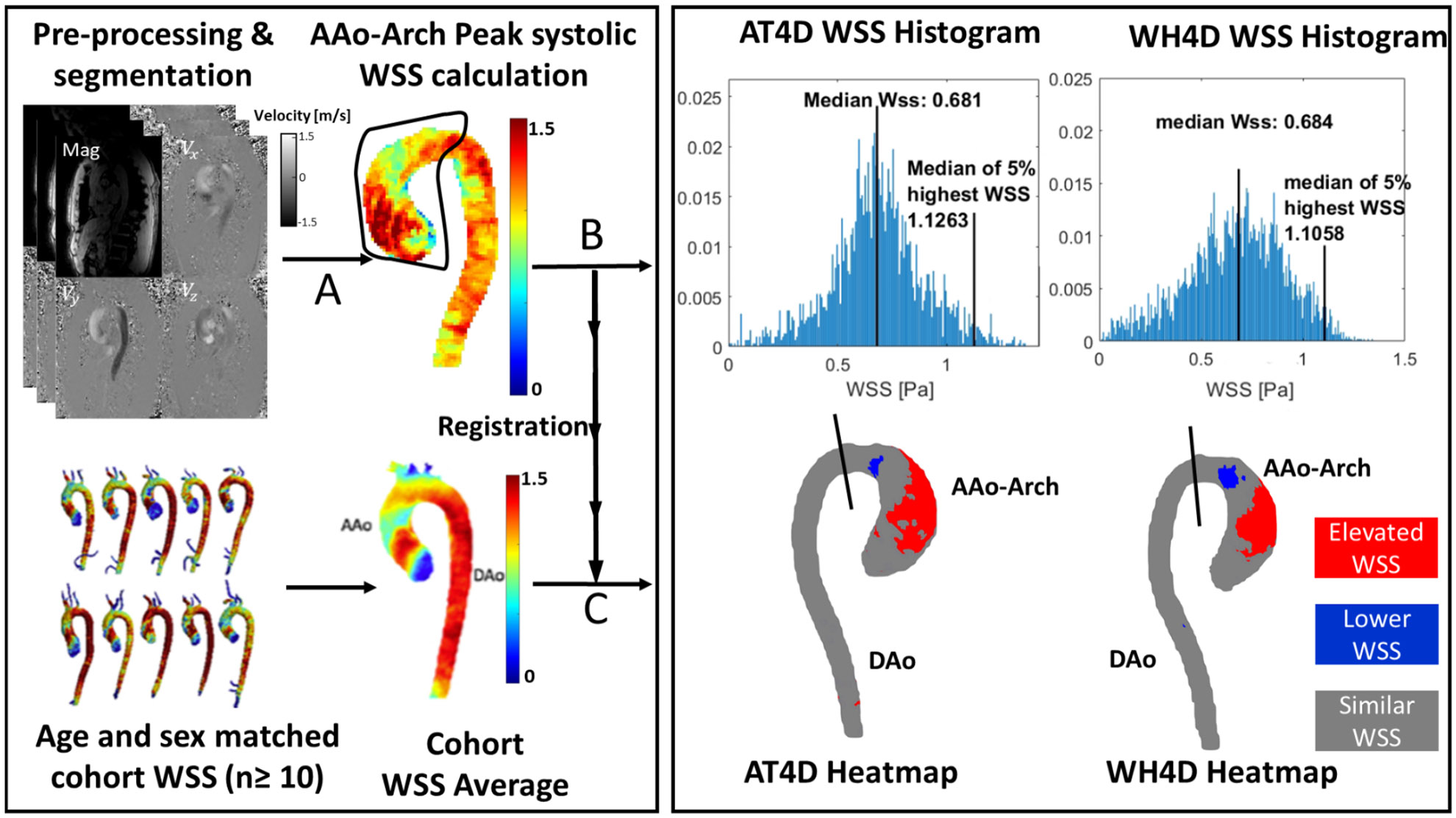
To evaluate the reproducibility of important biomarkers like wall shear stress (WSS), pulse wave velocity (PWV), and net flow across two 4D flow MRI imaging protocols with different coverages: aorta-targeted 4D flow MRI (AT4D) and whole-heart 4D flow (WH4D) protocols.

METHODS

Thirty-eight control subjects (43.2 ± 10.1 years old; 22 males) and ten patients (45.7 ± 8.9 years old; 7 males) with bicuspid aortic valve (BAV) were included. Each subject underwent AT4D and WH4D scans. Absolute WSS, PWV, and net flow were assessed for each patient across both protocols and compared using Bland-Altman analysis. Areas of elevated WSS were assessed for BAV patients across different WSS thresholds that define WSS to be elevated compared to a normal population average. A sensitivity analysis was conducted to determine the best WSS threshold at which WH4D-derived areas most closely resemble AT4D-derived areas. Inter-rater reproducibility was evaluated in twenty-four subjects.

RESULTS

AT4D and WH4D PWV and WSS estimates demonstrated good agreement (PWV: -0.12 ± 1.84 m/s, p = 0.4; Median WSS: 0.06 ± 0.13 Pa, p < 0.01; Maximum WSS: 0.04 ± 0.27 Pa, p = 0.07). Good agreement was also found for AAo net flow (8.14 ± 24.86 mL/cycle, p < 0.01). PWV correlated with age across protocols (AT4D: r = 0.68, p < 0.01; WH4D: r = 0.72, p < 0.01). Sensitivity analysis identified a WSS threshold where WH4D-derived areas of elevated WSS most closely resembled AT4D-derived areas. Inter-rater assessment of the tested parameters resulted in a small mean difference percentage of < 3%.

DATA CONCLUSION

PWV, WSS, and net flow demonstrated good agreement across protocols. The WSS threshold should be adjusted for WH4D estimates to optimally match AT4D-derived output. Reproducibility analysis showed good test-retest agreement. This study demonstrates the reproducibility of certain hemodynamic parameters across two 4D flow MRI protocol.

摘要

目的

通过两种不同覆盖范围的4D流MRI成像协议,即主动脉靶向4D流MRI(AT4D)和全心4D流(WH4D)协议,评估壁面剪应力(WSS)、脉搏波速度(PWV)和净流量等重要生物标志物的可重复性。

方法

纳入38名对照受试者(43.2±10.1岁;22名男性)和10名患有二叶式主动脉瓣(BAV)的患者(45.7±8.9岁;7名男性)。每位受试者均接受了AT4D和WH4D扫描。对每位患者在两种协议下的绝对WSS、PWV和净流量进行评估,并使用布兰德-奥特曼分析进行比较。针对BAV患者,在不同的WSS阈值下评估WSS升高区域,这些阈值定义了与正常人群平均值相比升高的WSS。进行敏感性分析以确定最佳WSS阈值,在该阈值下WH4D得出的区域与AT4D得出的区域最为相似。在24名受试者中评估了评分者间的可重复性。

结果

AT4D和WH4D的PWV和WSS估计值显示出良好的一致性(PWV:-0.12±1.84m/s,p = 0.4;中位WSS:0.06±0.13Pa,p < 0.01;最大WSS:0.04±0.27Pa,p = 0.07)。升主动脉净流量也显示出良好的一致性(8.14±24.86mL/周期,p < 0.01)。跨协议的PWV与年龄相关(AT4D:r = 0.68,p < 0.01;WH4D:r = 0.72,p < 0.01)。敏感性分析确定了一个WSS阈值,在该阈值下WH4D得出的WSS升高区域与AT4D得出的区域最为相似。对测试参数的评分者间评估导致平均差异百分比小于3%。

数据结论

PWV、WSS和净流量在各协议间显示出良好的一致性。对于WH4D估计值,应调整WSS阈值以使其与AT4D得出的输出最佳匹配。可重复性分析显示了良好的重测一致性。本研究证明了某些血流动力学参数在两种4D流MRI协议间的可重复性。

https://cdn.ncbi.nlm.nih.gov/pmc/blobs/22e6/11995425/69f9f80279fa/nihms-2069041-f0005.jpg
https://cdn.ncbi.nlm.nih.gov/pmc/blobs/22e6/11995425/bd1cc1016334/nihms-2069041-f0001.jpg
https://cdn.ncbi.nlm.nih.gov/pmc/blobs/22e6/11995425/112ecfc11033/nihms-2069041-f0002.jpg
https://cdn.ncbi.nlm.nih.gov/pmc/blobs/22e6/11995425/2d8f6f2af61d/nihms-2069041-f0003.jpg
https://cdn.ncbi.nlm.nih.gov/pmc/blobs/22e6/11995425/4bc7c030dcec/nihms-2069041-f0004.jpg
https://cdn.ncbi.nlm.nih.gov/pmc/blobs/22e6/11995425/69f9f80279fa/nihms-2069041-f0005.jpg