Gimbel Blake A, Roediger Donovan J, Anthony Mary E, Ernst Abigail M, Tuominen Kent A, Mueller Bryon A, de Water Erik, Rockhold Madeline N, Wozniak Jeffrey R
The Ohio State University and Nationwide Children's Hospital, United States.
University of Minnesota Twin Cities, United States.
Neuroimage Clin. 2025;45:103722. doi: 10.1016/j.nicl.2024.103722. Epub 2024 Dec 7.
To quantify regional subcortical brain volume anomalies in youth with fetal alcohol spectrum disorder (FASD), assess the relative sensitivity and specificity of abnormal volumes in FASD vs. a comparison group, and examine associations with cognitive function.
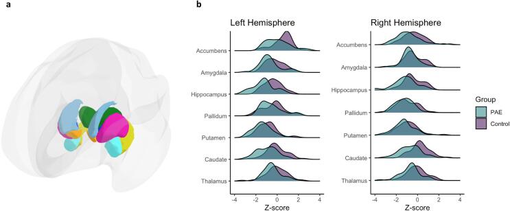
Participants: 47 children with FASD and 39 typically-developing comparison participants, ages 8-17 years, who completed physical evaluations, cognitive and behavioral testing, and an MRI brain scan. A large normative MRI dataset that controlled for sex, age, and intracranial volume was used to quantify the developmental status of 7 bilateral subcortical regional volumes. Z-scores were calculated based on volumetric differences from the normative sample. T-tests compared subcortical volumes across groups. Percentages of atypical volumes are reported as are sensitivity and specificity in discriminating groups. Lastly, Pearson correlations examined the relationships between subcortical volumes and neurocognitive performance.
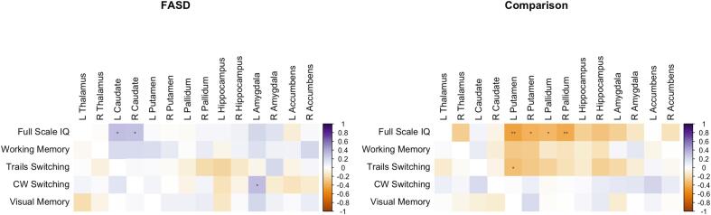
Participants with FASD demonstrated lower mean volumes across a majority of subcortical regions relative to the comparison group with prominent group differences in the bilateral hippocampi and bilateral caudate. More individuals with FASD (89%) had one or more abnormally small volume compared to 72% of the comparison group. The bilateral hippocampi, bilateral putamen, and right pallidum were most sensitive in discriminating those with FASD from the comparison group. Exploratory analyses revealed associations between subcortical volumes and cognitive functioning that differed across groups.
In this sample, youth with FASD had a greater number of atypically small subcortical volumes than individuals without FASD. Findings suggest MRI may have utility in identifying individuals with structural brain anomalies resulting from PAE.
量化患有胎儿酒精谱系障碍(FASD)的青少年的区域皮质下脑容量异常,评估FASD患者与对照组相比脑容量异常的相对敏感性和特异性,并研究其与认知功能的关联。
参与者:47名患有FASD的儿童和39名年龄在8至17岁之间发育正常的对照参与者,他们完成了身体评估、认知和行为测试以及脑部MRI扫描。使用一个控制了性别、年龄和颅内体积的大型MRI标准数据集来量化7个双侧皮质下区域体积的发育状况。根据与标准样本的体积差异计算Z分数。通过t检验比较各组之间的皮质下体积。报告非典型体积的百分比以及区分各组的敏感性和特异性。最后,通过Pearson相关性分析研究皮质下体积与神经认知表现之间的关系。
与对照组相比,患有FASD的参与者在大多数皮质下区域的平均体积较低,双侧海马体和双侧尾状核存在显著的组间差异。与对照组的72%相比,更多患有FASD的个体(89%)有一个或多个异常小的体积。双侧海马体、双侧壳核和右侧苍白球在区分FASD患者与对照组时最为敏感。探索性分析揭示了皮质下体积与认知功能之间的关联在不同组之间存在差异。
在这个样本中,患有FASD的青少年比没有FASD的个体有更多非典型的小皮质下体积。研究结果表明,MRI可能有助于识别因孕期酒精暴露导致的脑结构异常个体。