Wu Chengrong, Cai Hualei, Pu Qian, Yu Lei, Goswami Ashutosh, Mo Zhongyuan
Assisted Reproductive Center, Guiyang Women's and Children's Hospital (Guiyang Children's Hospital), Guiyang, 550000, China.
Department of Obstetrics and Gynecology, The Affiliated Hospital of Guizhou Medical University No. 28 of Guiyi Street, Yunyan District, Guiyang, Guizhou, 550000, China.
Open Med (Wars). 2024 Dec 4;19(1):20241077. doi: 10.1515/med-2024-1077. eCollection 2024.
Intrauterine adhesions (IUAs) are a significant clinical challenge, affecting reproductive health and leading to infertility or recurrent pregnancy loss. Understanding the molecular mechanisms underlying IUA prevention is crucial for developing effective treatment strategies.
To investigate the interaction between oviductal mucosal cells and endometrial cells and their effects on the expression of key molecules involved in embryo implantation, specifically leukemia inhibitory factor (LIF), avβ3, estrogen receptor (ER), and progesterone receptor (PR).
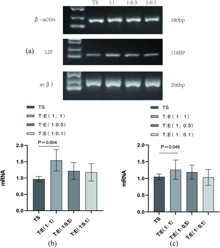
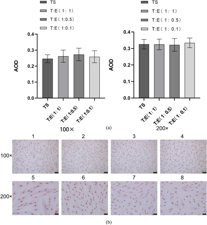
Tubal mucosa and endometrium specimens were collected from 22 patients undergoing surgical interventions. Cells were cultured alone and co-cultured at ratios of 1:1, 1:0.5, and 1:0.1. LIF, avβ3, ER, and PR expression levels were measured using real-time fluorescence quantitative polymerase chain reaction and enzyme-linked immunosorbent assay.
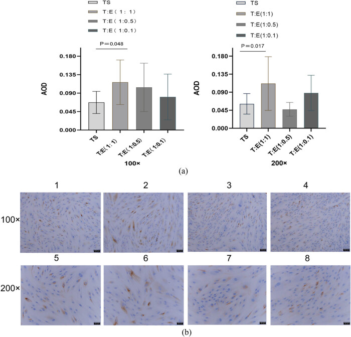
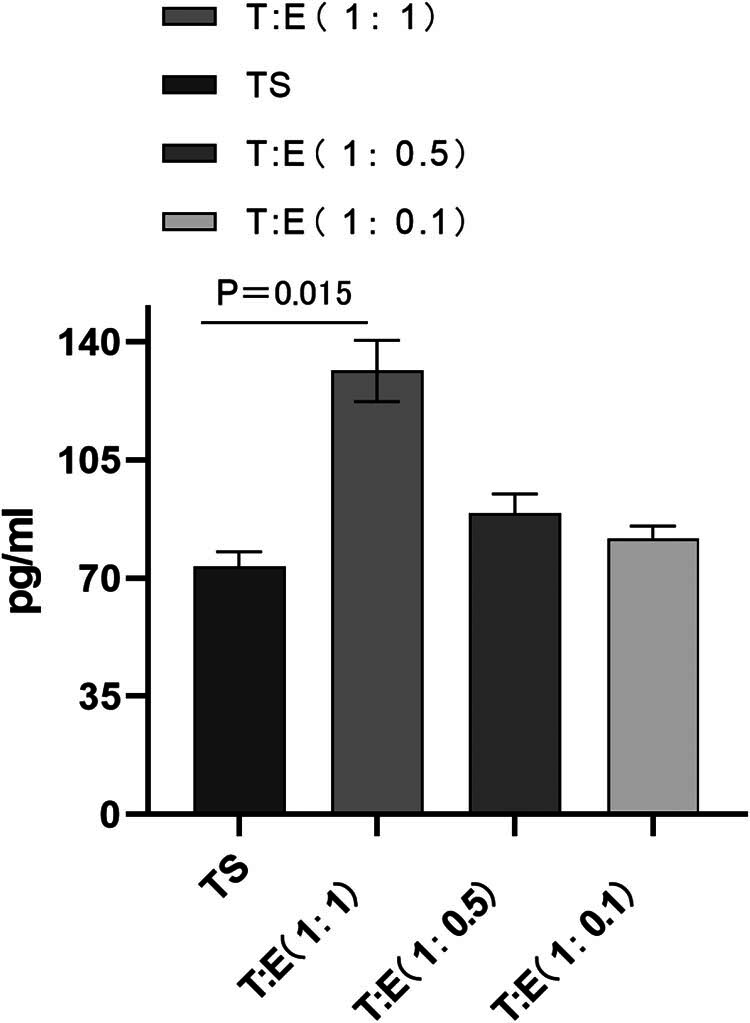
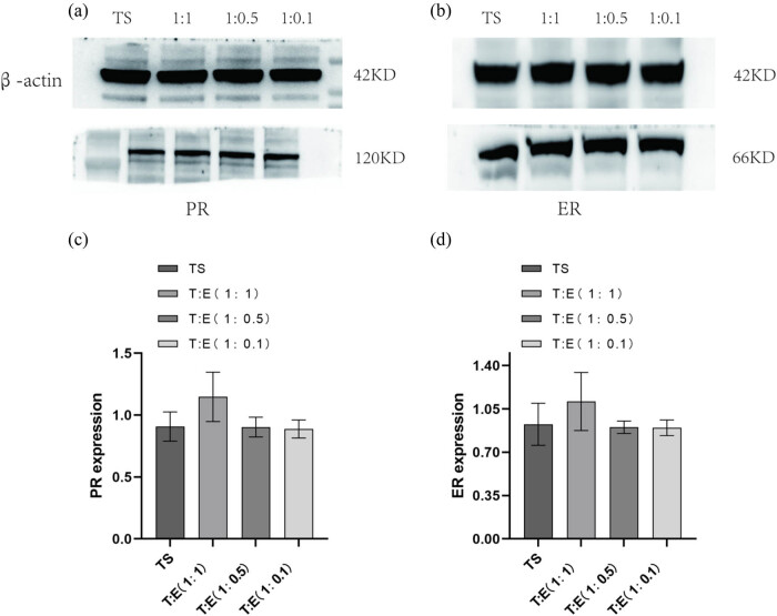
Our results demonstrated that LIF expression was significantly augmented in co-culture conditions, particularly in the 1:1 ratio, compared to oviductal mucosa monoculture ( < 0.05). Although LIF expression was also elevated in 1:0.5 and 1:0.1 co-culture ratios, these increases were not statistically significant ( > 0.05). For avβ3, increased expression was observed in the 1:1 co-culture group ( < 0.05), but no significant differences were detected in 1:0.5 and 1:0.1 co-culture groups. ER expression showed a downward trend in co-culture, but without statistical significance ( > 0.05), and PR expression remained stable across all groups.
Co-culture modulates key molecules involved in embryo implantation, particularly LIF and avβ3. These findings highlight the potential roles of LIF and avβ3 in IUA prevention strategies and provide important insights for future clinical interventions. Tubal mucosal cells can not only grow in the endometrial cell microenvironment, but also the tolerance of tubal mucosal cells can be improved when they are co-cultured.
宫腔粘连(IUAs)是一项重大的临床挑战,影响生殖健康并导致不孕或反复流产。了解宫腔粘连预防的分子机制对于制定有效的治疗策略至关重要。
研究输卵管黏膜细胞与子宫内膜细胞之间的相互作用及其对胚胎着床相关关键分子表达的影响,特别是白血病抑制因子(LIF)、αvβ3、雌激素受体(ER)和孕激素受体(PR)。
从22例接受手术干预的患者中收集输卵管黏膜和子宫内膜标本。细胞单独培养,并按1:1、1:0.5和1:0.1的比例共培养。使用实时荧光定量聚合酶链反应和酶联免疫吸附测定法测量LIF、αvβ3、ER和PR的表达水平。
我们的结果表明,与输卵管黏膜单独培养相比,共培养条件下LIF表达显著增加,尤其是在1:1比例时(P<0.05)。虽然在1:0.5和1:0.1共培养比例下LIF表达也有所升高,但这些增加无统计学意义(P>0.05)。对于αvβ3,在1:1共培养组中观察到表达增加(P<0.05),但在1:0.5和1:0.1共培养组中未检测到显著差异。共培养时ER表达呈下降趋势,但无统计学意义(P>0.05),且PR表达在所有组中保持稳定。
共培养可调节胚胎着床相关关键分子,特别是LIF和αvβ3。这些发现突出了LIF和αvβ3在宫腔粘连预防策略中的潜在作用,并为未来的临床干预提供了重要见解。输卵管黏膜细胞不仅能在子宫内膜细胞微环境中生长,而且共培养时其耐受性可得到改善。