• 文献检索
  • 文档翻译
  • 深度研究
  • 学术资讯
  • Suppr Zotero 插件Zotero 插件
  • 邀请有礼
  • 套餐&价格
  • 历史记录
应用&插件
Suppr Zotero 插件Zotero 插件浏览器插件Mac 客户端Windows 客户端微信小程序
定价
高级版会员购买积分包购买API积分包
服务
文献检索文档翻译深度研究API 文档MCP 服务
关于我们
关于 Suppr公司介绍联系我们用户协议隐私条款
关注我们

Suppr 超能文献

核心技术专利:CN118964589B侵权必究
粤ICP备2023148730 号-1Suppr @ 2026

文献检索

告别复杂PubMed语法,用中文像聊天一样搜索,搜遍4000万医学文献。AI智能推荐,让科研检索更轻松。

立即免费搜索

文件翻译

保留排版,准确专业,支持PDF/Word/PPT等文件格式,支持 12+语言互译。

免费翻译文档

深度研究

AI帮你快速写综述,25分钟生成高质量综述,智能提取关键信息,辅助科研写作。

立即免费体验

使用锥形束计算机断层扫描技术对印度亚人群下颌切牙的根管牙本质厚度和根管形态进行研究。

Radicular Dentin Thickness and Root Canal Morphology of Mandibular Incisors in Indian Subpopulation Using Cone Beam Computed Tomography.

作者信息

Krishnan Gokul, E Sudhanva M, Rangappa Anithakumari, Rangaswamy Vikram, Murthy Chethana S, Kumar N Naveen

机构信息

Department of Conservative Dentistry and Endodontics, Vokkaligara Sangha Dental College and Hospital, Bengaluru, IND.

出版信息

Cureus. 2024 Nov 9;16(11):e73355. doi: 10.7759/cureus.73355. eCollection 2024 Nov.

DOI:10.7759/cureus.73355
PMID:39659315
原文链接:https://pmc.ncbi.nlm.nih.gov/articles/PMC11628873/
Abstract

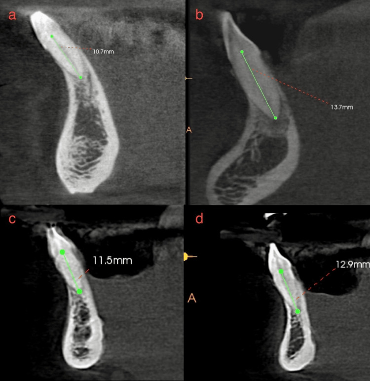
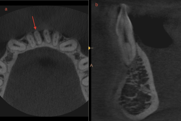
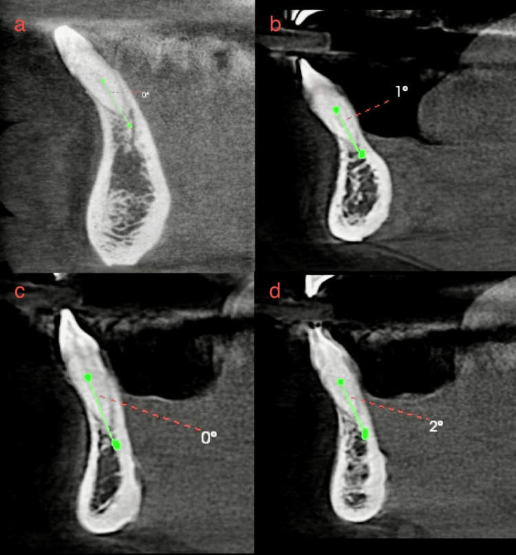
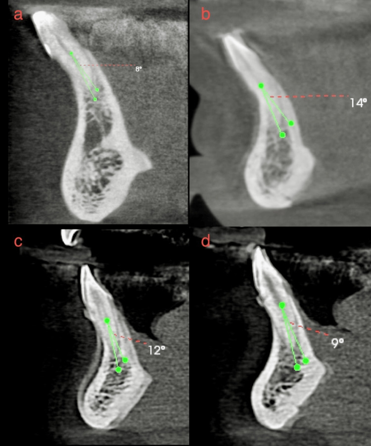
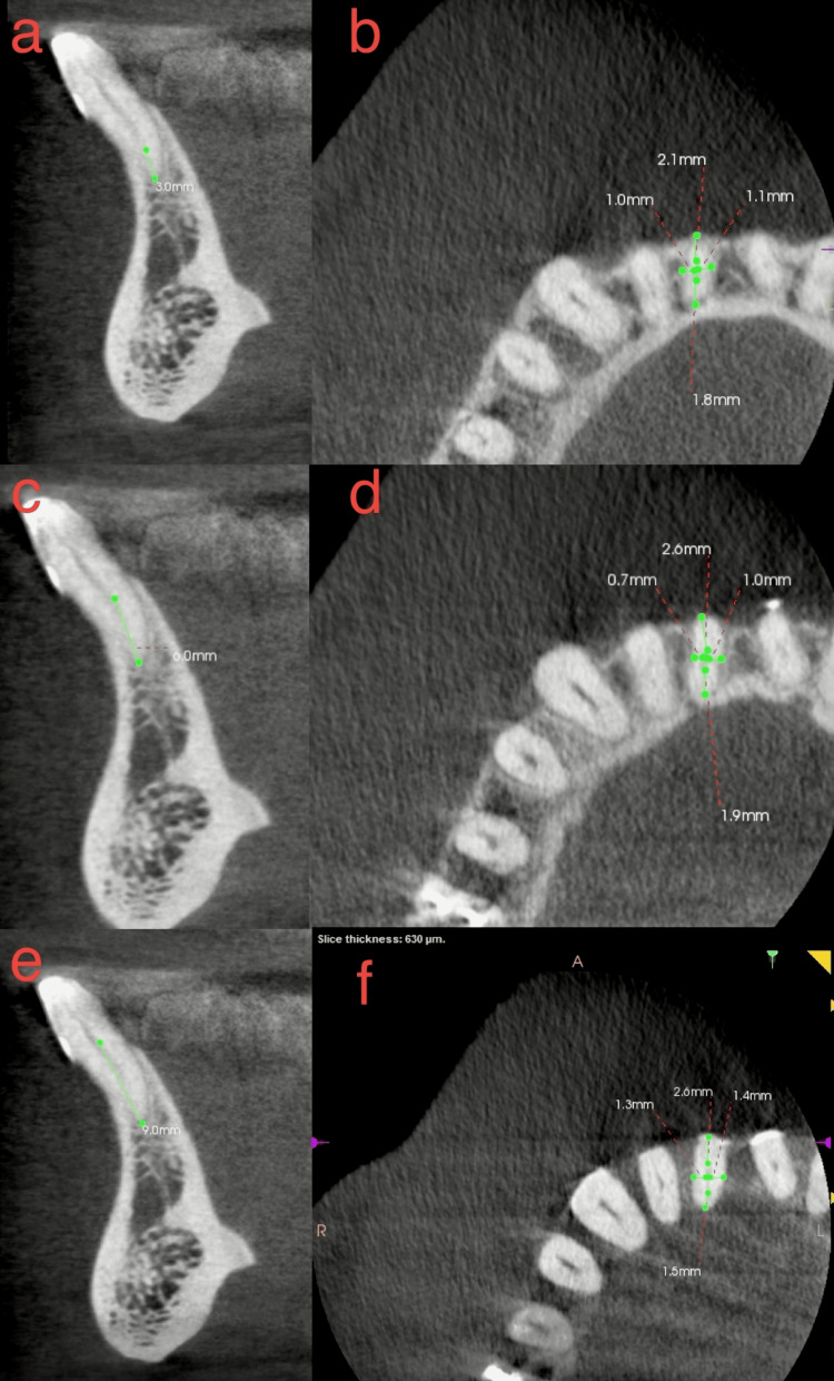
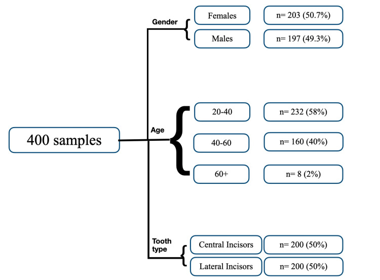
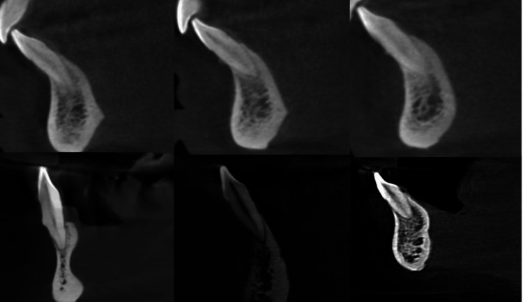
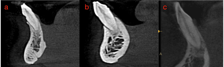
Aim Using two classifications, this study assessed root morphology and canal configuration and measured the Dentin thickness (DT) and canal shapes. Methods Cone beam computed tomography (CBCT) with 400 Mandibular Incisors was collected and assessed for the number, length, curvature of roots, number of canals, bifurcation level, configurations based on Vertucci's and Ahmed's classification, DT and canal shape at 3, 6, 9 mm from the apex. The collected data was subjected to statistical analysis with a level of significance at p<0.05. Results All samples had one root, averaging 12.769 ± 1.128 mm in central incisor (CI) and 13.044 ± 1.235 mm in lateral incisor (LI), with most roots being straight. Most samples had one canal in both teeth, with bifurcations most frequent in the middle third. The most frequent configuration was type 1 Vertucci or CI/LI by Ahmed, followed by type 3 or CI/LI. One sample, not classifiable under Vertucci, was classified as CI by Ahmed. The mean DT for CI was 3.18 ± 0.639 mm, 3.72 ± 0.671 mm and 4.43 ± 0.754 mm labiolingually and 1.578 ± 0.342 mm, 1.881 ± 0.374 mm, 2.283 ± 0.465 mm mesioditally at 3, 6, 9 mm from the apex, respectively. For LI, mean DT was 3.41 ± 0.916 mm, 3.90 ± 0.702 mm and 4.55 ± 0.746 mm labiolingually and 1.63 ± 0.322 mm, 1.981 ± 0.485 mm, 2.55 ± 0.470 mm mesioditally at 3, 6, 9 mm from the apex respectively, canal shape changed from oval to round, from apical to coronal. Conclusion Single canals were the most common, followed by two canals. The middle third of the canal had the most bifurcations. Vertucci type 1 or Ahmed's CI/LI  was the most commonly reported canal configuration, with one sample that could not be classified under Vertucci but could be classified using Ahmed classification. DT increased apical to coronal. The canal shape changed from oval to rounded, from apical to coronal.

摘要

目的 本研究使用两种分类方法评估牙根形态和根管形态,并测量牙本质厚度(DT)和根管形状。方法 收集400颗下颌切牙的锥形束计算机断层扫描(CBCT)图像,评估牙根的数量、长度、弯曲度、根管数量、分叉水平,根据Vertucci分类法和Ahmed分类法评估根管形态,测量根尖3、6、9 mm处的DT和根管形状。对收集的数据进行统计学分析,显著性水平为p<0.05。结果 所有样本均有一个牙根,中切牙(CI)平均长度为12.769±1.128 mm,侧切牙(LI)平均长度为13.044±1.235 mm,大多数牙根为直根。大多数样本的两颗牙均有一个根管,分叉最常见于中1/3处。最常见的形态为Vertucci分类法中的1型或Ahmed分类法中的CI/LI,其次是3型或CI/LI。有一个样本无法按照Vertucci分类法分类,但按照Ahmed分类法可归为CI。CI在根尖3、6、9 mm处的唇舌向平均DT分别为3.18±0.639 mm、3.72±0.671 mm和4.43±0.754 mm,近远中向平均DT分别为1.578±0.342 mm、1.881±0.374 mm和2.283±0.465 mm。LI在根尖3、6、9 mm处的唇舌向平均DT分别为3.41±0.916 mm、3.90±0.702 mm和4.55±0.746 mm,近远中向平均DT分别为1.63±0.322 mm、1.981±0.485 mm和2.55±0.470 mm,根管形状从根尖向冠方由椭圆形变为圆形。结论 单根管最为常见,其次是双根管。根管的中1/3处分叉最多。Vertucci 1型或Ahmed的CI/LI是最常报道的根管形态,有一个样本无法按照Vertucci分类法分类,但可按照Ahmed分类法分类。DT从根尖向冠方增加。根管形状从根尖向冠方由椭圆形变为圆形。

https://cdn.ncbi.nlm.nih.gov/pmc/blobs/efa4/11628873/0626b4260926/cureus-0016-00000073355-i08.jpg
https://cdn.ncbi.nlm.nih.gov/pmc/blobs/efa4/11628873/e2390a947b8c/cureus-0016-00000073355-i01.jpg
https://cdn.ncbi.nlm.nih.gov/pmc/blobs/efa4/11628873/54b389764d4f/cureus-0016-00000073355-i02.jpg
https://cdn.ncbi.nlm.nih.gov/pmc/blobs/efa4/11628873/7d7ca5e011b5/cureus-0016-00000073355-i03.jpg
https://cdn.ncbi.nlm.nih.gov/pmc/blobs/efa4/11628873/1350c01c8089/cureus-0016-00000073355-i04.jpg
https://cdn.ncbi.nlm.nih.gov/pmc/blobs/efa4/11628873/6cd93334a34c/cureus-0016-00000073355-i05.jpg
https://cdn.ncbi.nlm.nih.gov/pmc/blobs/efa4/11628873/b7ddf303b5c1/cureus-0016-00000073355-i06.jpg
https://cdn.ncbi.nlm.nih.gov/pmc/blobs/efa4/11628873/f70dcdded177/cureus-0016-00000073355-i07.jpg
https://cdn.ncbi.nlm.nih.gov/pmc/blobs/efa4/11628873/0626b4260926/cureus-0016-00000073355-i08.jpg

相似文献

1
Radicular Dentin Thickness and Root Canal Morphology of Mandibular Incisors in Indian Subpopulation Using Cone Beam Computed Tomography.使用锥形束计算机断层扫描技术对印度亚人群下颌切牙的根管牙本质厚度和根管形态进行研究。
Cureus. 2024 Nov 9;16(11):e73355. doi: 10.7759/cureus.73355. eCollection 2024 Nov.
2
Root canal morphology of mandibular anterior permanent teeth in Turkish sub-population using two classification systems: a cone-beam computed tomography study.使用两种分类系统对土耳其亚人群下颌前恒牙根管形态的锥形束计算机断层扫描研究
Odontology. 2025 Apr;113(2):799-808. doi: 10.1007/s10266-024-01000-5. Epub 2024 Sep 16.
3
Cone Beam Computed Tomography Study of Root Canal Morphology of Permanent Mandibular Incisors in Indian Subpopulation.印度亚人群下颌恒切牙根管形态的锥形束计算机断层扫描研究
Pol J Radiol. 2017 Jul 7;82:371-375. doi: 10.12659/PJR.901840. eCollection 2017.
4
Comparative study of Vertucci and Ahmed classifications to evaluate the main root canal configuration of mandibular incisors in a Brazilian population.比较Vertucci分类法和Ahmed分类法以评估巴西人群下颌切牙的主要根管形态
Aust Endod J. 2022 Dec;48(3):409-414. doi: 10.1111/aej.12576. Epub 2021 Oct 9.
5
Cone-beam Computed Tomography Analysis of the Root Canal Morphology of Mandibular Incisors Using Two Classification Systems in a Spanish Subpopulation: A Cross-Sectional Study.使用两种分类系统对西班牙亚人群下颌切牙根管形态的锥形束计算机断层扫描分析:一项横断面研究。
Eur Endod J. 2024 Mar 8;9(2):106 - 113. doi: 10.14744/eej.2023.10327. Epub 2024 Feb 21.
6
Evaluation of the root and canal morphology of mandibular permanent anterior teeth in an Iranian population by cone-beam computed tomography.通过锥形束计算机断层扫描评估伊朗人群下颌恒前牙的根管形态。
J Dent (Tehran). 2013 May;10(4):358-66. Epub 2013 May 31.
7
Root canal morphology and bifurcating/merging sites of permanent mandibular incisors in 9- to 12-year-old children: An in-vivo 3D visualization analysis in China.9至12岁儿童恒牙下颌切牙的根管形态及分叉/融合部位:中国的一项体内三维可视化分析
Arch Oral Biol. 2023 Nov;155:105781. doi: 10.1016/j.archoralbio.2023.105781. Epub 2023 Aug 5.
8
Root and Root Canal Anatomy of Primary Mandibular Central Incisor, Lateral Incisor, and Canine in Indian Children: A Cone Beam Computed Tomography Study.印度儿童下颌乳中切牙、侧切牙和尖牙的牙根及根管解剖:一项锥形束计算机断层扫描研究
Int J Dent. 2022 Mar 7;2022:7191134. doi: 10.1155/2022/7191134. eCollection 2022.
9
Vertucci's root canal configuration of 11,376 mandibular anteriors and its relationship with distolingual roots in mandibular first molars in a Cantonese population: a cone-beam computed tomography study.一项针对广东人群下颌第一磨牙近中舌根管与远中舌根管及 11,376 颗下颌前牙根管形态的锥形束 CT 研究
BMC Oral Health. 2022 Apr 16;22(1):130. doi: 10.1186/s12903-022-02078-5.
10
Evaluation of root and root canal morphology of mandibular and maxillary canine teeth in Turkish subpopulation by cone beam computed tomography with using two classification systems.使用两种分类系统,通过锥形束计算机断层扫描评估土耳其亚人群下颌和上颌尖牙的牙根及根管形态。
BMC Oral Health. 2024 Dec 18;24(1):1499. doi: 10.1186/s12903-024-05252-z.

本文引用的文献

1
Cone-beam Computed Tomography Analysis of the Root Canal Morphology of Mandibular Incisors Using Two Classification Systems in a Spanish Subpopulation: A Cross-Sectional Study.使用两种分类系统对西班牙亚人群下颌切牙根管形态的锥形束计算机断层扫描分析:一项横断面研究。
Eur Endod J. 2024 Mar 8;9(2):106 - 113. doi: 10.14744/eej.2023.10327. Epub 2024 Feb 21.
2
Root canal morphology of anterior permanent teeth in Jordanian population using two classification systems: a cone-beam computed tomography study.使用两种分类系统对约旦人群的前恒牙根管形态进行锥形束 CT 研究。
BMC Oral Health. 2024 Feb 2;24(1):170. doi: 10.1186/s12903-024-03934-2.
3
Endodontic Management of Mandibular anterior teeth and premolars with Vertucci's Type VIII canal morphology: A Rare Case.
下颌前牙和前磨牙Vertucci VIII型根管形态的牙髓治疗:1例罕见病例
J Conserv Dent. 2022 Mar-Apr;25(2):197-201. doi: 10.4103/jcd.jcd_518_21. Epub 2022 May 4.
4
Evaluation of root canal morphology in permanent maxillary and mandibular anterior teeth in Saudi subpopulation using two classification systems: a CBCT study.利用两种分类系统评估沙特人群中上颌和下颌前恒牙的根管形态:CBCT 研究。
BMC Oral Health. 2022 May 10;22(1):171. doi: 10.1186/s12903-022-02187-1.
5
Prevalence of Missed Canals and Their Association with Apical Periodontitis in Posterior Endodontically Treated Teeth: A CBCT Study.根管遗漏的患病率及其与后牙根管治疗后患牙根尖周炎的相关性:一项CBCT研究
Int J Dent. 2021 Jun 28;2021:9962429. doi: 10.1155/2021/9962429. eCollection 2021.
6
Root and canal morphology of the anterior permanent dentition in Malaysian population using two classification systems: A CBCT clinical study.马来西亚人群前永久恒牙的根和根管形态:两种分类系统的 CBCT 临床研究。
Aust Endod J. 2021 Aug;47(2):202-216. doi: 10.1111/aej.12454. Epub 2020 Nov 7.
7
Dentin thickness as a risk factor for vertical root fracture in endodontically treated teeth: a case-control study.牙本质厚度作为根管治疗后牙齿发生垂直根裂的风险因素:一项病例对照研究。
Clin Oral Investig. 2021 Mar;25(3):1099-1105. doi: 10.1007/s00784-020-03406-1. Epub 2020 Jun 26.
8
Cone-beam-computed-tomography of the symmetry of root canal anatomy in mandibular incisors.下颌切牙根管解剖对称性的锥形束计算机断层扫描
J Oral Sci. 2020;62(2):180-183. doi: 10.2334/josnusd.19-0113.
9
A study of the symmetry of roots and root canal morphology in mandibular anterior teeth using cone-beam computed tomographic imaging in a Polish population.应用锥形束 CT 成像技术研究波兰人群下颌前牙根管形态和根尖的对称性。
Folia Morphol (Warsz). 2020;79(4):835-844. doi: 10.5603/FM.a2019.0128. Epub 2019 Dec 5.
10
Anatomical analysis of permanent mandibular incisors in a Saudi Arabian population: An cone-beam computed tomography study.沙特阿拉伯人群下颌恒切牙的解剖学分析:一项锥形束计算机断层扫描研究。
Niger J Clin Pract. 2019 Nov;22(11):1611-1616. doi: 10.4103/njcp.njcp_291_19.