• 文献检索
  • 文档翻译
  • 深度研究
  • 学术资讯
  • Suppr Zotero 插件Zotero 插件
  • 邀请有礼
  • 套餐&价格
  • 历史记录
应用&插件
Suppr Zotero 插件Zotero 插件浏览器插件Mac 客户端Windows 客户端微信小程序
定价
高级版会员购买积分包购买API积分包
服务
文献检索文档翻译深度研究API 文档MCP 服务
关于我们
关于 Suppr公司介绍联系我们用户协议隐私条款
关注我们

Suppr 超能文献

核心技术专利:CN118964589B侵权必究
粤ICP备2023148730 号-1Suppr @ 2026

文献检索

告别复杂PubMed语法,用中文像聊天一样搜索,搜遍4000万医学文献。AI智能推荐,让科研检索更轻松。

立即免费搜索

文件翻译

保留排版,准确专业,支持PDF/Word/PPT等文件格式,支持 12+语言互译。

免费翻译文档

深度研究

AI帮你快速写综述,25分钟生成高质量综述,智能提取关键信息,辅助科研写作。

立即免费体验

使用两种分类系统对约旦人群的前恒牙根管形态进行锥形束 CT 研究。

Root canal morphology of anterior permanent teeth in Jordanian population using two classification systems: a cone-beam computed tomography study.

机构信息

Department of Conservative Dentistry, Faculty of Dentistry, Jordan University of Science and Technology, Box 3030, Irbid, Jordan.

Dental Teaching Clinics, Jordan University of Science and Technology, Irbid, Jordan.

出版信息

BMC Oral Health. 2024 Feb 2;24(1):170. doi: 10.1186/s12903-024-03934-2.

DOI:10.1186/s12903-024-03934-2
PMID:38308267
原文链接:https://pmc.ncbi.nlm.nih.gov/articles/PMC10837869/
Abstract

BACKGROUND

Adequate knowledge of root canal morphology and its variation is essential for success of root canal treatment and to overcome treatemnt failure. The aim of this study was to investigate the root and canal morphology of mandibular anterior teeth using 2 classification systems.

METHODS

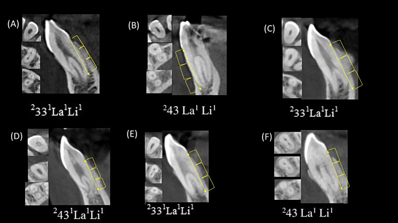
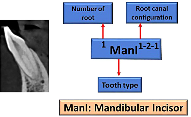
3342 lower anteriors were evaluated from 557 CBCT scans. The images were examined in sagittal, axial and coronal views using a CS 3D imaging software (V3.10.4, Carestream Dental). Demographic data recorded, the number of roots and canal's morphology were described according to Vertucci and Ahmed classifications.

RESULTS

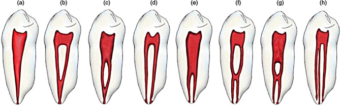
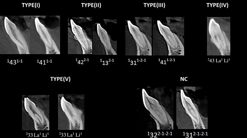
Frequency of Type I configuration was significantly the highest in incisors and canines (76%, N = 2539), followed by Type III (20.6%, N = 687). Type II (1.1%, N = 37), IV (1.1%, N = 37), and V (0.3%, N = 11) were rarely encountered. 0.9% (N = 31) of the teeth could not be classified with the Vertucci System. The frequency of 2 roots (MA in Ahmed classification) which has no correspondence in the Vertucci classification, was 1.1% (N = 38), it was significantly higher in canines and in females (35 canines and 3 laterals). A moderate correlation in root canal morpology was found between the left and right sides (V > 0.30). 80% (N = 2538) of the teeth did not exhibit any divergence/merging. The bifurcation level occurred mostly in the middle third of the root.

CONCLUSIONS

One fourth of anterior teeth had variation from the simple type I canal configuration and therefore requires attention during treatment. The new classification system offers a more accurate and simplified presentation of canal morphology.

CLINICAL RELEVANCE

The prevalence and mid root bifurcation of second canal in lower anteriors requires attention to ensure adequate quality root canal treatment without compromising the integrity of teeth.

摘要

背景

充分了解根管形态及其变异对于根管治疗的成功和克服治疗失败至关重要。本研究旨在使用两种分类系统研究下颌前牙的根管和根形态。

方法

从 557 个 CBCT 扫描中评估了 3342 个下颌前牙。使用 CS 3D 成像软件(V3.10.4,Carestream Dental)在矢状面、轴面和冠状面检查图像。记录人口统计学数据,根据 Vertucci 和 Ahmed 分类描述根管的数量和形态。

结果

在切牙和尖牙中,I 型构型的频率明显最高(76%,N=2539),其次是 III 型(20.6%,N=687)。II 型(1.1%,N=37)、IV 型(1.1%,N=37)和 V 型(0.3%,N=11)很少见。0.9%(N=31)的牙齿无法用 Vertucci 系统进行分类。在 Ahmed 分类中没有对应 Vertucci 分类的双根(MA)的频率为 1.1%(N=38),在尖牙和女性中明显更高(35 颗尖牙和 3 颗侧切牙)。左右两侧根管形态存在中度相关性(V>0.30)。80%(N=2538)的牙齿没有任何分叉/融合。分叉发生在根的中三分之一处。

结论

四分之一的前牙的根管形态与简单的 I 型不同,因此在治疗过程中需要注意。新的分类系统提供了一种更准确和简化的根管形态呈现方式。

临床意义

下颌前牙的第二根管的存在和中根分叉需要注意,以确保高质量的根管治疗,同时不损害牙齿的完整性。

https://cdn.ncbi.nlm.nih.gov/pmc/blobs/316b/10837869/8bbebc804aab/12903_2024_3934_Fig4_HTML.jpg
https://cdn.ncbi.nlm.nih.gov/pmc/blobs/316b/10837869/9723a776f9e3/12903_2024_3934_Fig1_HTML.jpg
https://cdn.ncbi.nlm.nih.gov/pmc/blobs/316b/10837869/e324b8fa5b0b/12903_2024_3934_Fig2_HTML.jpg
https://cdn.ncbi.nlm.nih.gov/pmc/blobs/316b/10837869/e8976d594836/12903_2024_3934_Fig3_HTML.jpg
https://cdn.ncbi.nlm.nih.gov/pmc/blobs/316b/10837869/8bbebc804aab/12903_2024_3934_Fig4_HTML.jpg

相似文献

1
Root canal morphology of anterior permanent teeth in Jordanian population using two classification systems: a cone-beam computed tomography study.使用两种分类系统对约旦人群的前恒牙根管形态进行锥形束 CT 研究。
BMC Oral Health. 2024 Feb 2;24(1):170. doi: 10.1186/s12903-024-03934-2.
2
Evaluation of root canal morphology in permanent maxillary and mandibular anterior teeth in Saudi subpopulation using two classification systems: a CBCT study.利用两种分类系统评估沙特人群中上颌和下颌前恒牙的根管形态:CBCT 研究。
BMC Oral Health. 2022 May 10;22(1):171. doi: 10.1186/s12903-022-02187-1.
3
Root and canal morphology of the anterior permanent dentition in Malaysian population using two classification systems: A CBCT clinical study.马来西亚人群前永久恒牙的根和根管形态:两种分类系统的 CBCT 临床研究。
Aust Endod J. 2021 Aug;47(2):202-216. doi: 10.1111/aej.12454. Epub 2020 Nov 7.
4
Root and canal morphology of the permanent anterior dentition in a Black South African population using cone-beam computed tomography and two classification systems.使用锥形束计算机断层扫描和两种分类系统研究南非黑人永久性前牙的根管和牙根形态。
J Oral Sci. 2022 Jul 1;64(3):218-223. doi: 10.2334/josnusd.22-0027. Epub 2022 May 30.
5
Vertucci's root canal configuration of 11,376 mandibular anteriors and its relationship with distolingual roots in mandibular first molars in a Cantonese population: a cone-beam computed tomography study.一项针对广东人群下颌第一磨牙近中舌根管与远中舌根管及 11,376 颗下颌前牙根管形态的锥形束 CT 研究
BMC Oral Health. 2022 Apr 16;22(1):130. doi: 10.1186/s12903-022-02078-5.
6
Prevalence of Isthmi and Root Canal Configurations in Mandibular Permanent Teeth Using Cone-Beam Computed Tomography.下颌恒磨牙应用锥形束 CT 研究其根管和峡部的解剖形态。
Biomed Res Int. 2024 Jul 31;2024:9969860. doi: 10.1155/2024/9969860. eCollection 2024.
7
Root canal morphology of mandibular anterior permanent teeth in Turkish sub-population using two classification systems: a cone-beam computed tomography study.使用两种分类系统对土耳其亚人群下颌前恒牙根管形态的锥形束计算机断层扫描研究
Odontology. 2025 Apr;113(2):799-808. doi: 10.1007/s10266-024-01000-5. Epub 2024 Sep 16.
8
Root canal configuration of human permanent anterior teeth: A cross-sectional study using cone-beam computed tomography in the Pakistani population.巴基斯坦人群中应用锥形束 CT 对人恒前牙根管形态的横断面研究。
J Pak Med Assoc. 2024 Sep;74(9):1608-1612. doi: 10.47391/JPMA.10199.
9
Assessment of the root and canal morphology in the permanent dentition of Saudi Arabian population using cone beam computed and micro-computed tomography - a systematic review.使用锥形束计算机断层扫描和微计算机断层扫描评估沙特阿拉伯人群恒牙的根管和根管形态:系统评价。
BMC Oral Health. 2024 Mar 16;24(1):343. doi: 10.1186/s12903-024-04101-3.
10
Root canal morphology of permanent teeth in a Malaysian subpopulation using cone-beam computed tomography.马来西亚人群恒牙根尖形态的锥形束 CT 研究。
BMC Oral Health. 2019 Jan 14;19(1):14. doi: 10.1186/s12903-019-0710-z.

引用本文的文献

1
Evaluation of root and root canal morphology in maxillary first molar teeth: A cone-beam computed tomography study using two classification systems in a Japanese population.上颌第一磨牙牙根及根管形态的评估:一项在日本人群中使用两种分类系统的锥形束计算机断层扫描研究。
J Dent Sci. 2025 Jul;20(3):1554-1561. doi: 10.1016/j.jds.2024.12.005. Epub 2024 Dec 14.
2
Comparative study of Vertucci's and Ahmed's classifications to evaluate the root canal configuration of mandibular incisors in Saudi Arabian subpopulation.比较Vertucci分类法和Ahmed分类法以评估沙特阿拉伯亚人群下颌切牙的根管形态。
Pak J Med Sci. 2025 Jun;41(6):1658-1663. doi: 10.12669/pjms.41.6.11972.
3

本文引用的文献

1
Critical analysis of a new system to classify root and canal morphology - A systematic review.根管形态分类新系统的批判性分析——系统评价。
Aust Endod J. 2023 Dec;49(3):750-768. doi: 10.1111/aej.12780. Epub 2023 Sep 8.
2
Assessment of the Prevalence of Radix Entomolaris and Distolingual Canal in Mandibular First Molars in 15 Countries: A Multinational Cross-sectional Study with Meta-analysis.评估 15 个国家下颌第一磨牙的齿根(Radix Entomolaris)和远舌侧管(Distolingual Canal)的流行情况:一项多国横断面研究的荟萃分析。
J Endod. 2023 Oct;49(10):1308-1318. doi: 10.1016/j.joen.2023.06.011. Epub 2023 Jun 30.
3
Root and canal configurations of maxillary first premolars in 22 countries using two classification systems: a multinational cross-sectional study.
使用两种分类系统对22个国家上颌第一前磨牙的根管形态进行的多国家横断面研究。
Sci Rep. 2025 Jun 2;15(1):19290. doi: 10.1038/s41598-025-02669-6.
4
Evaluation of root and root canal morphology in maxillary premolar teeth: A cone-beam computed tomography study using two classification systems in a Japanese population.上颌前磨牙牙根及根管形态的评估:一项在日本人群中使用两种分类系统的锥形束计算机断层扫描研究。
J Dent Sci. 2025 Apr;20(2):927-935. doi: 10.1016/j.jds.2024.08.024. Epub 2024 Sep 8.
5
Micro-computed tomographic evaluation of root and canal anatomy of maxillary first premolars in Iraqi sub-population.伊拉克亚人群上颌第一前磨牙牙根及根管解剖结构的显微计算机断层扫描评估
Sci Rep. 2025 Mar 28;15(1):10821. doi: 10.1038/s41598-025-94590-1.
6
Evaluation of root and root canal morphology of mandibular and maxillary canine teeth in Turkish subpopulation by cone beam computed tomography with using two classification systems.使用两种分类系统,通过锥形束计算机断层扫描评估土耳其亚人群下颌和上颌尖牙的牙根及根管形态。
BMC Oral Health. 2024 Dec 18;24(1):1499. doi: 10.1186/s12903-024-05252-z.
7
Radicular Dentin Thickness and Root Canal Morphology of Mandibular Incisors in Indian Subpopulation Using Cone Beam Computed Tomography.使用锥形束计算机断层扫描技术对印度亚人群下颌切牙的根管牙本质厚度和根管形态进行研究。
Cureus. 2024 Nov 9;16(11):e73355. doi: 10.7759/cureus.73355. eCollection 2024 Nov.
8
Classifying the internal anatomy of anterior teeth in the Yemeni population using two systems: a retrospective CBCT study.使用两种系统对也门人群前牙的内部解剖结构进行分类:一项回顾性CBCT研究。
Odontology. 2025 Jan;113(1):416-431. doi: 10.1007/s10266-024-00965-7. Epub 2024 Jun 27.
Prediction of root canal lengths and pulp volume of the maxillary permanent first molar based on stature, crown diameters, and facial morphometry.
基于身高、冠径和面型测量预测上颌恒第一磨牙的根管长度和牙髓容积。
Anat Sci Int. 2023 Jul;98(3):454-462. doi: 10.1007/s12565-023-00727-5. Epub 2023 Apr 20.
4
Assessment of the Prevalence of Middle Mesial Canal in Mandibular First Molar: A Multinational Cross-sectional Study with Meta-analysis.下颌第一磨牙近中中根管患病率的评估:一项荟萃分析的多国横断面研究
J Endod. 2023 May;49(5):549-558. doi: 10.1016/j.joen.2023.02.012. Epub 2023 Mar 1.
5
Evaluation of root canal morphology in permanent maxillary and mandibular anterior teeth in Saudi subpopulation using two classification systems: a CBCT study.利用两种分类系统评估沙特人群中上颌和下颌前恒牙的根管形态:CBCT 研究。
BMC Oral Health. 2022 May 10;22(1):171. doi: 10.1186/s12903-022-02187-1.
6
Comparative study of Vertucci and Ahmed classifications to evaluate the main root canal configuration of mandibular incisors in a Brazilian population.比较Vertucci分类法和Ahmed分类法以评估巴西人群下颌切牙的主要根管形态
Aust Endod J. 2022 Dec;48(3):409-414. doi: 10.1111/aej.12576. Epub 2021 Oct 9.
7
Root and canal morphology of the anterior permanent dentition in Malaysian population using two classification systems: A CBCT clinical study.马来西亚人群前永久恒牙的根和根管形态:两种分类系统的 CBCT 临床研究。
Aust Endod J. 2021 Aug;47(2):202-216. doi: 10.1111/aej.12454. Epub 2020 Nov 7.
8
Application of a new system for classifying root canal morphology in undergraduate teaching and clinical practice: a national survey in Malaysia.在本科教学和临床实践中应用根管形态分类新系统:马来西亚全国调查。
Int Endod J. 2020 Jun;53(6):871-879. doi: 10.1111/iej.13271. Epub 2020 Mar 16.
9
Root and canal configurations of maxillary premolars in a South African subpopulation using cone beam computed tomography and two classification systems.使用锥形束计算机断层扫描和两种分类系统对南非亚人群上颌前磨牙的根管形态进行研究
J Oral Sci. 2020;62(1):93-97. doi: 10.2334/josnusd.19-0160.
10
Evaluation of Root Canal Anatomy in Mandibular Incisors Using CBCT Imaging Technique in an Iranian Population.在伊朗人群中使用CBCT成像技术评估下颌切牙的根管解剖结构
J Dent (Shiraz). 2019 Mar;20(1):24-29. doi: 10.30476/DENTJODS.2019.44559.