Doran Michael, Grant John, Soussahn Samer
Department of Orthopaedic Surgery, University of Michigan, Ann Arbor, MI, USA.
Department of Radiology, University of Michigan, Ann Arbor, MI, USA.
Radiol Case Rep. 2024 Nov 28;20(2):1034-1040. doi: 10.1016/j.radcr.2024.10.148. eCollection 2025 Feb.
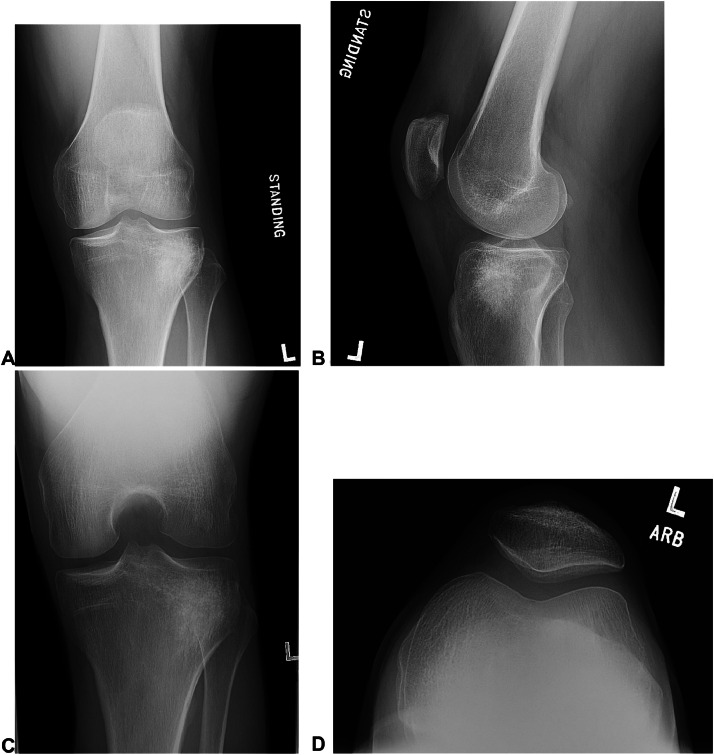
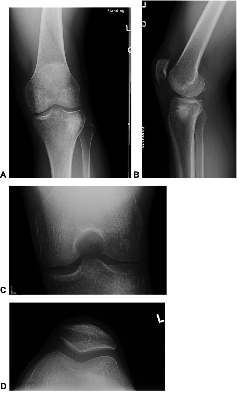
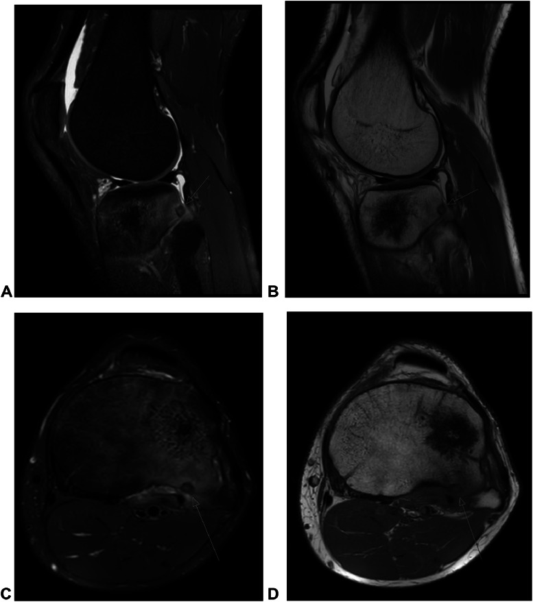
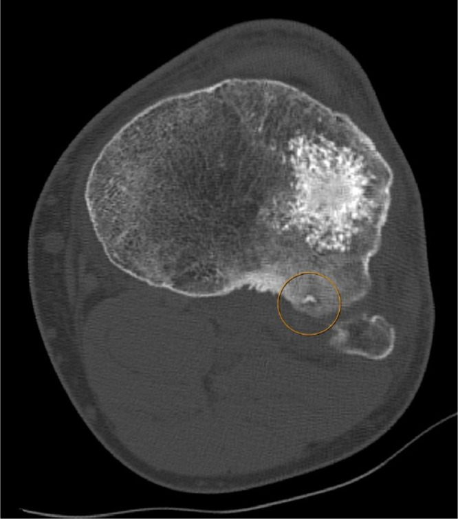
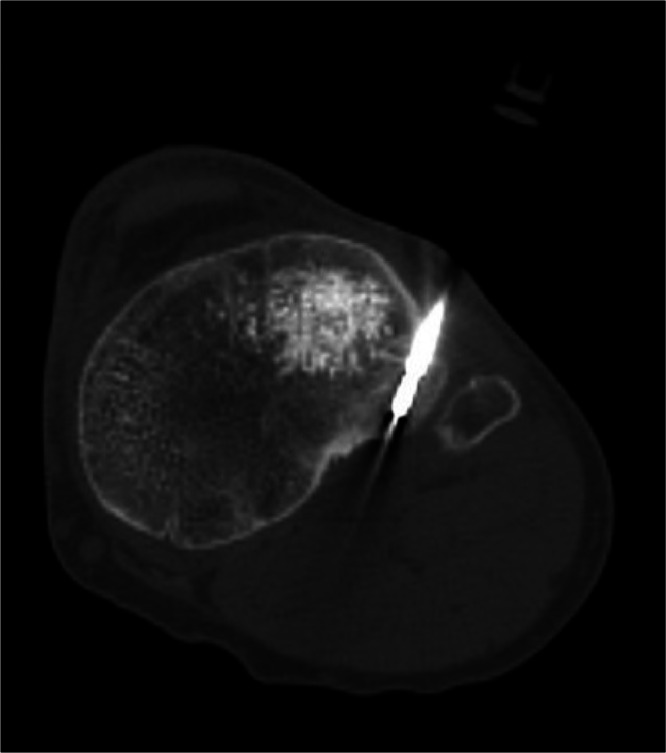
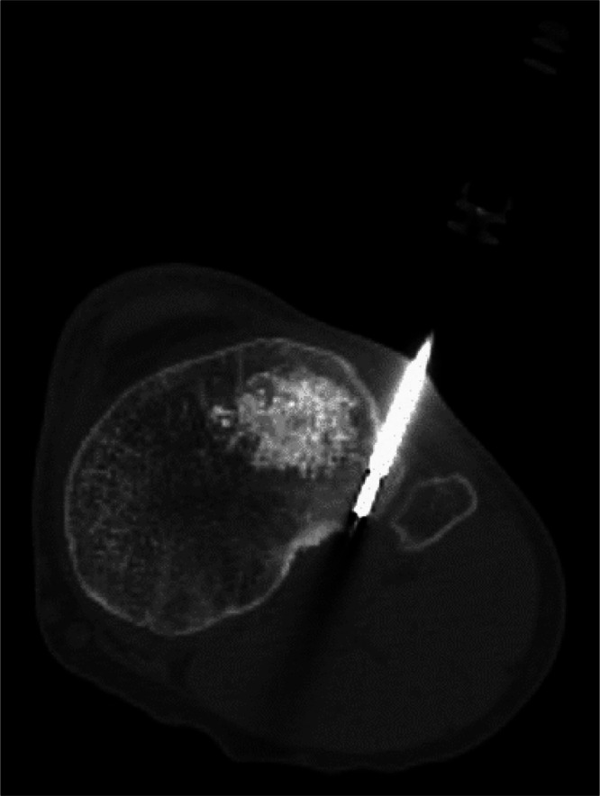
Osteoid Osteoma (OO) is a common primary bone tumor that often presents with night pain in younger orthopedic patients. Although typically extra-articular, intra-articular presentations may be difficult to diagnose. While magnetic resonance imaging (MRI) provides excellent detailed imaging of the articular surface, it has been reported to lead to occasional misdiagnosis given limitations in spatial resolution, particularly for smaller lesions. Computed tomography (CT) remains the gold standard imaging modality for OO. The treatment for osteoid osteoma consists of medical management, minimally invasive image guided techniques, and surgical resection in order of most conservative to most aggressive. We present the case of a 31-year-old male with persistent posterolateral knee pain after subchondroplasty. CT demonstrated an OO in the posterior tibial plateau. The patient was successfully treated with CT-guided percutaneous radiofrequency ablation with complete resolution of symptoms. We also provide a brief literature review of the diagnosis and treatment of OO to help heighten the awareness of this sometimes inconspicuous diagnosis.
骨样骨瘤(OO)是一种常见的原发性骨肿瘤,在年轻的骨科患者中常表现为夜间疼痛。虽然通常位于关节外,但关节内表现可能难以诊断。虽然磁共振成像(MRI)能提供出色的关节表面详细成像,但据报道,由于空间分辨率的限制,偶尔会导致误诊,尤其是对于较小的病变。计算机断层扫描(CT)仍然是骨样骨瘤的金标准成像方式。骨样骨瘤的治疗包括药物治疗、微创影像引导技术和手术切除,从最保守到最激进依次进行。我们报告一例31岁男性患者,在软骨下成形术后持续存在膝关节后外侧疼痛。CT显示胫骨后平台有一个骨样骨瘤。该患者通过CT引导下经皮射频消融成功治疗,症状完全缓解。我们还对骨样骨瘤的诊断和治疗进行了简要的文献综述,以帮助提高对这种有时不明显诊断的认识。