Oteiza-Rius Inés, Estenaga Pérez de Albéniz Ángela, Hernández-Martin Ángela, Alfageme Roldán Fernando, García-Martínez Francisco Javier
Dermatology Department, Clínica Universidad de Navarra, Pamplona, Spain.
Dermatology Department, Hospital Universitario de Navarra, Pamplona, Spain.
J Ultrasound Med. 2025 Apr;44(4):703-709. doi: 10.1002/jum.16632. Epub 2024 Dec 12.
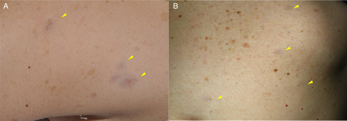
Red-blue neurofibromas (RBNs), found in up to 29% of adult neurofibromatosis type 1 (NF1) patients, present as red-blue macules measuring 1-2 cm in diameter, primarily on the trunk. Despite their prevalence, RBNs often go unnoticed due to their subtle appearance. Ultrasound characterization serves as a diagnostic clue yet lacks comprehensive studies in both adult and pediatric populations. This study aims to define and compare RBNs' prevalence, characteristics, and ultrasound features in adult and pediatric patients with NF1.
This prospective study involved 118 patients (92 pediatric patients and 26 adults) diagnosed with NF1. Clinical examinations combined with cutaneous ultrasound scans using linear multifrequency probes (L4-12t, L10-22, ML6-15, or L8-18 MHz) were performed in order to determine the prevalence, and clinical and sonographic characteristics of RBN in both populations. Statistical analyses were performed using t tests and chi-square tests.
RBNs were found in 26.3% (31) of the patients after clinical examination, including 179 lesions. RBN prevalence differed significantly between pediatric (10.9%) and adult (66.7%) patients. Lesions were primarily on the trunk and exhibited similar clinical characteristics. Ultrasound reveals RBNs as hypoechoic, oval lesions with irregular borders. Our results show that pediatric RBNs are typically more superficial and hypoechogenic, while adult RBNs are deeper and more heterogeneous.
Ultrasound findings showed subtle differences in lesion depth, morphology, and echogenicity between these 2 age-related groups. These changes highlight ultrasound's role in identifying RBNs in patients with NF1 and monitoring their evolution.
红蓝色神经纤维瘤(RBNs)在高达29%的成年1型神经纤维瘤病(NF1)患者中出现,表现为直径1 - 2厘米的红蓝色斑疹,主要位于躯干。尽管其发病率较高,但由于外观不明显,RBNs常常未被注意到。超声特征可作为诊断线索,但在成人和儿童群体中均缺乏全面研究。本研究旨在确定并比较成人和儿童NF1患者中RBNs的患病率、特征及超声表现。
这项前瞻性研究纳入了118例诊断为NF1的患者(92例儿童患者和26例成人患者)。进行临床检查并结合使用线性多频探头(L4 - 12t、L10 - 22、ML6 - 15或L8 - 18 MHz)进行皮肤超声扫描,以确定两组人群中RBNs的患病率、临床及超声特征。使用t检验和卡方检验进行统计分析。
临床检查后,26.3%(31例)的患者发现有RBNs,共179个病灶。儿童(10.9%)和成人(66.7%)患者的RBNs患病率差异显著。病灶主要位于躯干,且具有相似的临床特征。超声显示RBNs为边界不规则的低回声椭圆形病灶。我们的结果表明,儿童RBNs通常更表浅且回声更低,而成人RBNs更深且更不均匀。
超声检查结果显示,这两个与年龄相关的组在病灶深度、形态和回声方面存在细微差异。这些变化突出了超声在识别NF1患者中的RBNs及其病情演变监测中的作用。