• 文献检索
  • 文档翻译
  • 深度研究
  • 学术资讯
  • Suppr Zotero 插件Zotero 插件
  • 邀请有礼
  • 套餐&价格
  • 历史记录
应用&插件
Suppr Zotero 插件Zotero 插件浏览器插件Mac 客户端Windows 客户端微信小程序
定价
高级版会员购买积分包购买API积分包
服务
文献检索文档翻译深度研究API 文档MCP 服务
关于我们
关于 Suppr公司介绍联系我们用户协议隐私条款
关注我们

Suppr 超能文献

核心技术专利:CN118964589B侵权必究
粤ICP备2023148730 号-1Suppr @ 2026

文献检索

告别复杂PubMed语法,用中文像聊天一样搜索,搜遍4000万医学文献。AI智能推荐,让科研检索更轻松。

立即免费搜索

文件翻译

保留排版,准确专业,支持PDF/Word/PPT等文件格式,支持 12+语言互译。

免费翻译文档

深度研究

AI帮你快速写综述,25分钟生成高质量综述,智能提取关键信息,辅助科研写作。

立即免费体验

口腔正畸临床摄影技术

Techniques for Capturing Clinical Photographs in Orthodontics.

作者信息

Shahrul Al Imran, Mohd Shukor Nabilla, Norman Noraina Hafizan

机构信息

Department of Family Oral Health, Universiti Kebangsaan Malaysia, Kuala Lumpur, MYS.

Department of Orthodontics, Management and Science University (MSU) Medical Centre, Shah Alam, MYS.

出版信息

Cureus. 2024 Nov 13;16(11):e73629. doi: 10.7759/cureus.73629. eCollection 2024 Nov.

DOI:10.7759/cureus.73629
PMID:39677078
原文链接:https://pmc.ncbi.nlm.nih.gov/articles/PMC11645108/
Abstract

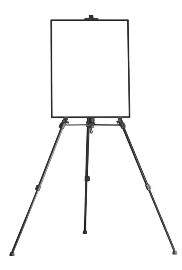
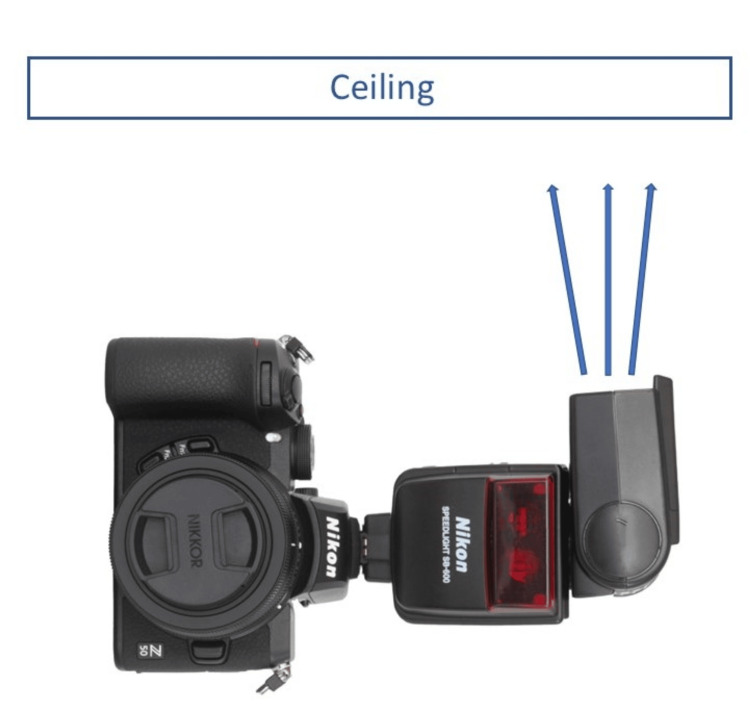
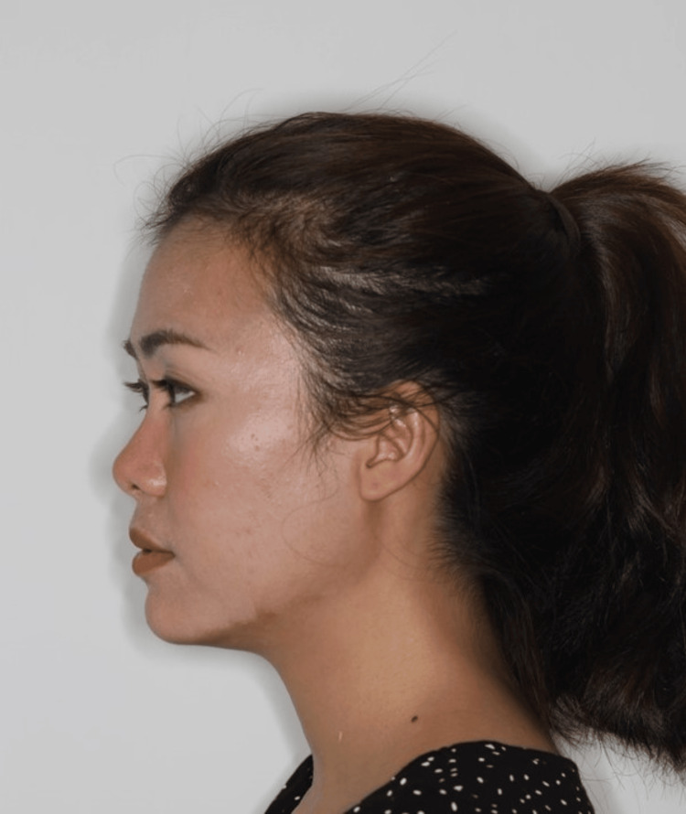
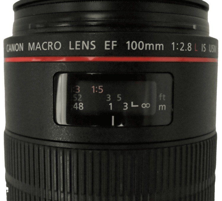
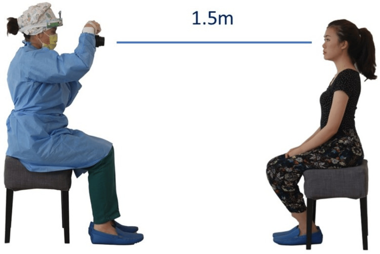
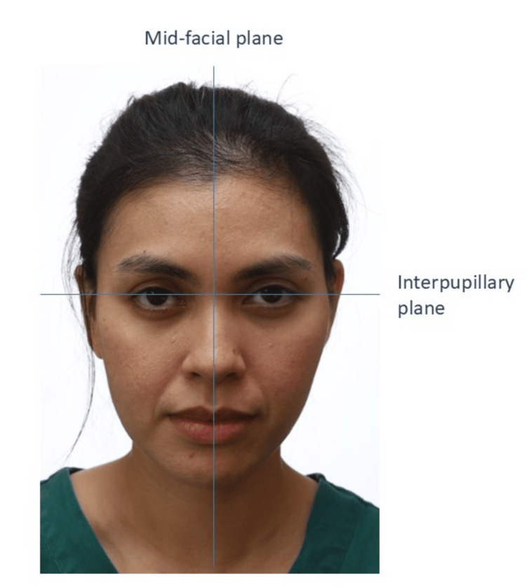
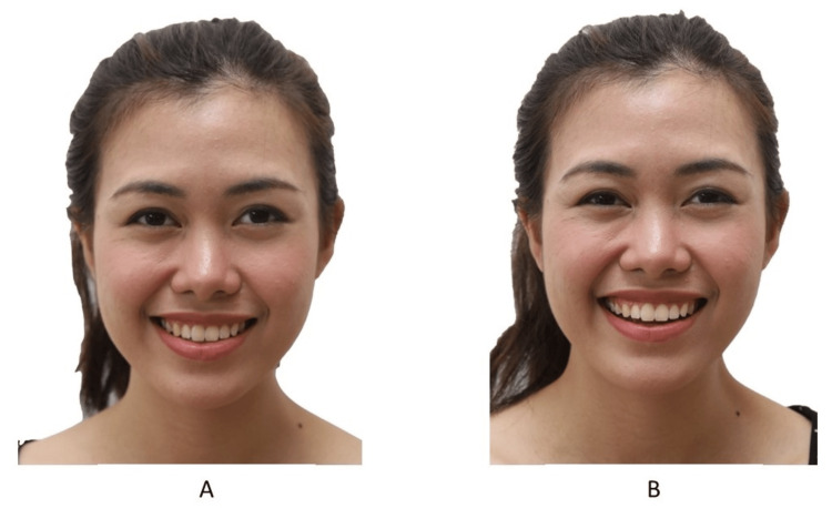
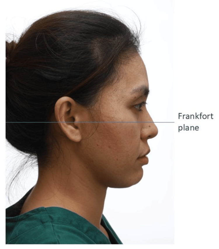
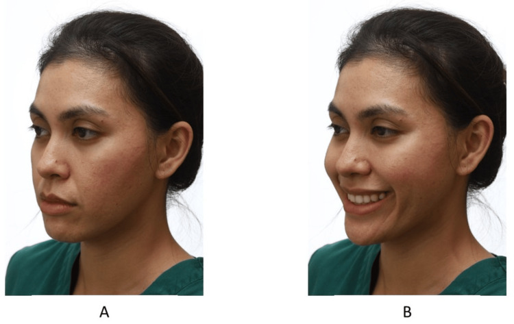
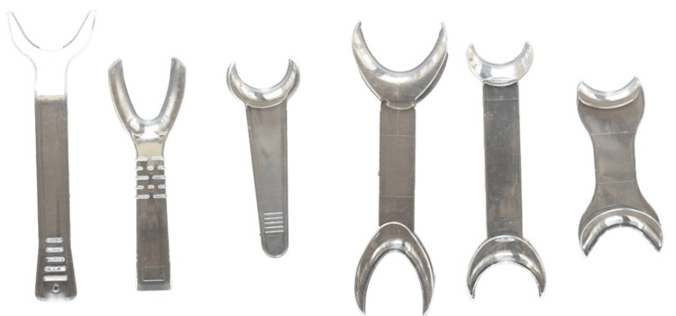
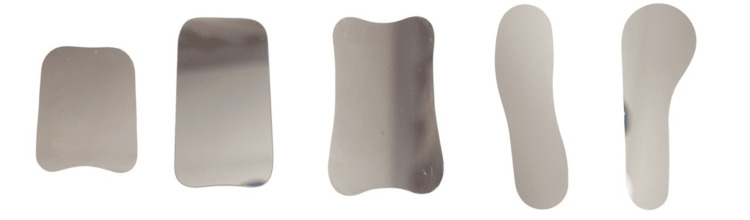
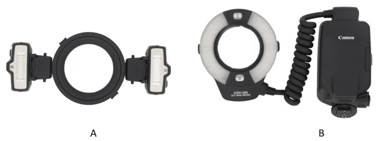
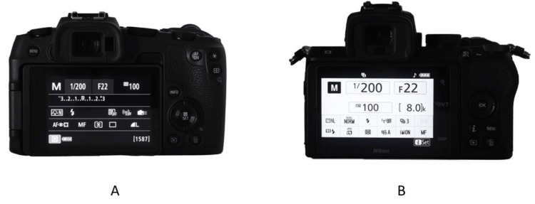
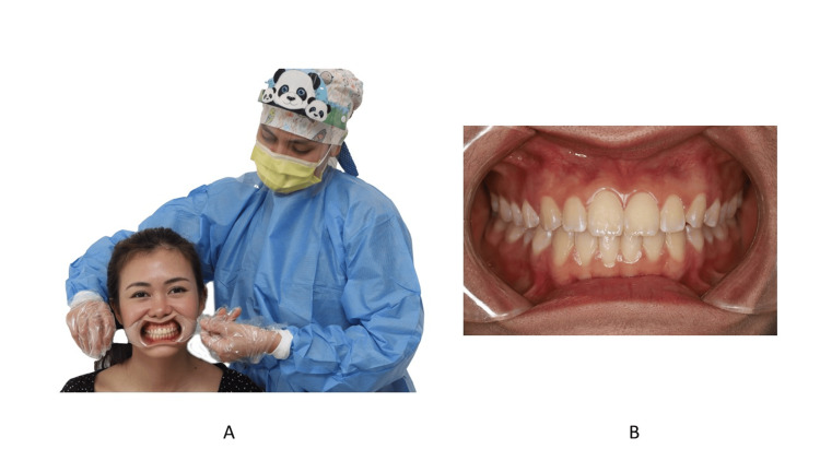
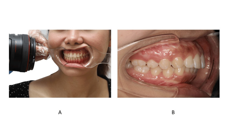
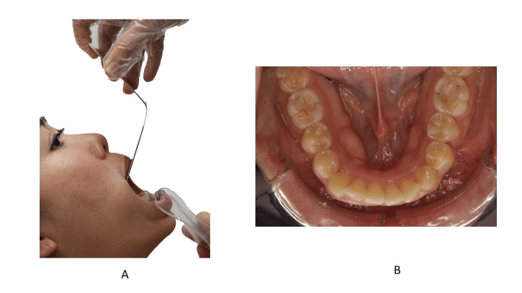
High-quality clinical photography is essential in orthodontics, playing a crucial role in diagnosis, treatment planning, patient education, and professional communication. However, capturing consistently clear and detailed orthodontic photographs can be challenging, particularly without knowledge of standardized techniques and equipment. This article provides a comprehensive guide on the key elements of effective orthodontic photography, including camera settings, positioning, and lighting. Illustrating each step in the process aims to equip orthodontists with the skills needed to achieve high-quality, reproducible clinical photos that enhance the quality of patient care and support clinical documentation.

摘要

高质量的临床摄影在正畸学中至关重要,在诊断、治疗计划、患者教育和专业交流中发挥着关键作用。然而,要始终拍摄出清晰、详细的正畸照片可能具有挑战性,尤其是在不了解标准化技术和设备的情况下。本文提供了一份关于有效正畸摄影关键要素的全面指南,包括相机设置、定位和照明。对过程中的每一步进行说明旨在使正畸医生具备获得高质量、可重复的临床照片所需的技能,从而提高患者护理质量并支持临床记录。

https://cdn.ncbi.nlm.nih.gov/pmc/blobs/c443/11645108/1f739646884a/cureus-0016-00000073629-i22.jpg
https://cdn.ncbi.nlm.nih.gov/pmc/blobs/c443/11645108/62850ae463de/cureus-0016-00000073629-i01.jpg
https://cdn.ncbi.nlm.nih.gov/pmc/blobs/c443/11645108/f70276be0e6b/cureus-0016-00000073629-i02.jpg
https://cdn.ncbi.nlm.nih.gov/pmc/blobs/c443/11645108/dcc066f33881/cureus-0016-00000073629-i03.jpg
https://cdn.ncbi.nlm.nih.gov/pmc/blobs/c443/11645108/b19510a55c9a/cureus-0016-00000073629-i04.jpg
https://cdn.ncbi.nlm.nih.gov/pmc/blobs/c443/11645108/ce860082f5f0/cureus-0016-00000073629-i05.jpg
https://cdn.ncbi.nlm.nih.gov/pmc/blobs/c443/11645108/f0a84bb02b9b/cureus-0016-00000073629-i06.jpg
https://cdn.ncbi.nlm.nih.gov/pmc/blobs/c443/11645108/ca07d7d0d4e8/cureus-0016-00000073629-i07.jpg
https://cdn.ncbi.nlm.nih.gov/pmc/blobs/c443/11645108/fd55af503283/cureus-0016-00000073629-i08.jpg
https://cdn.ncbi.nlm.nih.gov/pmc/blobs/c443/11645108/494904ddfc05/cureus-0016-00000073629-i09.jpg
https://cdn.ncbi.nlm.nih.gov/pmc/blobs/c443/11645108/eef5c1362cf4/cureus-0016-00000073629-i10.jpg
https://cdn.ncbi.nlm.nih.gov/pmc/blobs/c443/11645108/652184e10711/cureus-0016-00000073629-i11.jpg
https://cdn.ncbi.nlm.nih.gov/pmc/blobs/c443/11645108/232e4a6cd1f3/cureus-0016-00000073629-i12.jpg
https://cdn.ncbi.nlm.nih.gov/pmc/blobs/c443/11645108/2549343a5839/cureus-0016-00000073629-i13.jpg
https://cdn.ncbi.nlm.nih.gov/pmc/blobs/c443/11645108/677950d857d3/cureus-0016-00000073629-i14.jpg
https://cdn.ncbi.nlm.nih.gov/pmc/blobs/c443/11645108/66fd83bd6fb9/cureus-0016-00000073629-i15.jpg
https://cdn.ncbi.nlm.nih.gov/pmc/blobs/c443/11645108/827921ec90ab/cureus-0016-00000073629-i16.jpg
https://cdn.ncbi.nlm.nih.gov/pmc/blobs/c443/11645108/86f3ee62f1d0/cureus-0016-00000073629-i17.jpg
https://cdn.ncbi.nlm.nih.gov/pmc/blobs/c443/11645108/846a271f1297/cureus-0016-00000073629-i18.jpg
https://cdn.ncbi.nlm.nih.gov/pmc/blobs/c443/11645108/d419ea8a22f6/cureus-0016-00000073629-i19.jpg
https://cdn.ncbi.nlm.nih.gov/pmc/blobs/c443/11645108/98ac77757641/cureus-0016-00000073629-i20.jpg
https://cdn.ncbi.nlm.nih.gov/pmc/blobs/c443/11645108/7d1c768df72e/cureus-0016-00000073629-i21.jpg
https://cdn.ncbi.nlm.nih.gov/pmc/blobs/c443/11645108/1f739646884a/cureus-0016-00000073629-i22.jpg

相似文献

1
Techniques for Capturing Clinical Photographs in Orthodontics.口腔正畸临床摄影技术
Cureus. 2024 Nov 13;16(11):e73629. doi: 10.7759/cureus.73629. eCollection 2024 Nov.
2
Quality of clinical photographs taken by orthodontists, professional photographers, and orthodontic auxiliaries.正畸医生、专业摄影师和正畸辅助人员所拍摄临床照片的质量。
Am J Orthod Dentofacial Orthop. 2009 May;135(5):657-62. doi: 10.1016/j.ajodo.2007.04.038.
3
Clinical Photography in Orthodontic Practice: Insights from a Nationwide Survey in Spain.正畸临床实践中的临床摄影:来自西班牙全国性调查的见解。
J Clin Med. 2025 Mar 14;14(6):1984. doi: 10.3390/jcm14061984.
4
The establishment of the photo management system for orthodontics and its clinical application.正畸照片管理系统的建立及其临床应用。
Int J Comput Dent. 2005 Apr;8(2):161-8.
5
Diagnostic accuracy of intraoral photographic orthodontic records.口腔摄影正畸记录的诊断准确性。
J Esthet Restor Dent. 2019 Jan;31(1):64-71. doi: 10.1111/jerd.12426. Epub 2018 Oct 31.
6
A Standardized Approach to Extra-Oral and Intra-Oral Digital Photography.口腔外和口腔内数字摄影的标准化方法。
J Vis Exp. 2022 Jul 22(185). doi: 10.3791/63627.
7
[Documentation of orthodontic clinical cases. Photography in orthodontics. 2].[正畸临床病例记录。正畸摄影。2]
Mondo Ortod. 1989 Mar-Apr;14(2):127-44.
8
The Reliability and Validity of Intraoral Photographs in Assessing Orthodontic Treatment Need.口内照片在评估正畸治疗需求中的信度和效度
Orthod Craniofac Res. 2025 Jun;28(3):474-484. doi: 10.1111/ocr.12896. Epub 2025 Jan 13.
9
Comparison of 10 digital SLR cameras for orthodontic photography.10款数码单反相机用于正畸摄影的比较。
J Orthod. 2006 Sep;33(3):223-30. doi: 10.1179/146531205225021687.
10
Dynamic Video-Photograph Studio: A New Rhinoplasty Documentation.动态视频摄影工作室:一种新的隆鼻术记录方法。
Aesthetic Plast Surg. 2020 Jun;44(3):1093-1096. doi: 10.1007/s00266-020-01673-7. Epub 2020 Mar 20.

本文引用的文献

1
Mirrorless cameras in orthodontic practice.无反光镜相机在正畸治疗中的应用。
J Orthod. 2021 Sep;48(3):295-299. doi: 10.1177/14653125211000055. Epub 2021 Mar 24.
2
Diagnostic accuracy of intraoral photographic orthodontic records.口腔摄影正畸记录的诊断准确性。
J Esthet Restor Dent. 2019 Jan;31(1):64-71. doi: 10.1111/jerd.12426. Epub 2018 Oct 31.
3
The best choice of equipment to obtain high quality standardised results in intra-oral photography - a comparison between the common practice in the UK and the gold standard set by the literature.
在口腔内摄影中获得高质量标准化结果的最佳设备选择——英国的常见做法与文献设定的黄金标准之比较
J Vis Commun Med. 2018 Apr;41(2):90-96. doi: 10.1080/17453054.2018.1451250. Epub 2018 May 1.
4
Clinical Dental Photography in Orthodontic Practice.正畸临床中的牙科摄影
Prim Dent J. 2016 Nov 1;5(4):38-44. doi: 10.1308/205016816820209488.
5
Clinical photographs: the gold standard, an update.临床照片:金标准,更新。
Prog Orthod. 2012 Nov;13(3):296-303. doi: 10.1016/j.pio.2011.12.002. Epub 2012 Mar 21.
6
History and current use of clinical photography in orthodontics.正畸临床摄影的历史与当前应用
J Calif Dent Assoc. 2009 Mar;37(3):173-4.
7
Digital dental photography. Part 8: intra-oral set-ups.数字牙科摄影。第8部分:口腔内设置。
Br Dent J. 2009 Aug 22;207(4):151-7. doi: 10.1038/sj.bdj.2009.715.
8
Digital dental photography. Part 7: extra-oral set-ups.数字牙科摄影。第7部分:口外设置。
Br Dent J. 2009 Aug 8;207(3):103-10. doi: 10.1038/sj.bdj.2009.667.
9
Digital dental photography. Part 6: camera settings.数字牙科摄影。第6部分:相机设置。
Br Dent J. 2009 Jul 25;207(2):63-9. doi: 10.1038/sj.bdj.2009.607.
10
Digital dental photography. Part 5: lighting.数字牙科摄影。第5部分:照明。
Br Dent J. 2009 Jul 11;207(1):13-8. doi: 10.1038/sj.bdj.2009.558.