Wang Xiao-Jie, Zhou Jia-Ping, Pan Yao, Yu Ri-Sheng
Department of Radiology, Second Affiliated Hospital, Zhejiang University School of Medicine, Hangzhou, China.
Front Oncol. 2024 Nov 29;14:1463362. doi: 10.3389/fonc.2024.1463362. eCollection 2024.
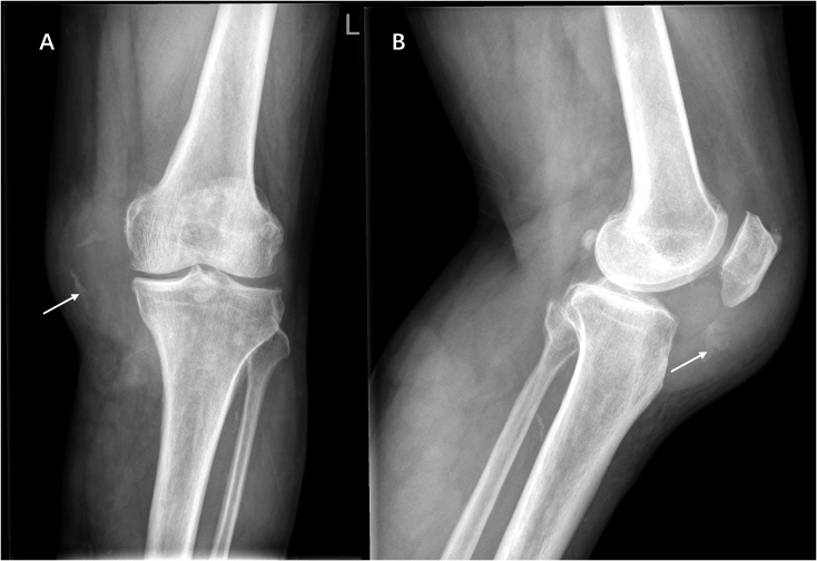
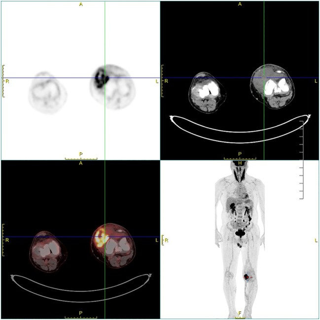
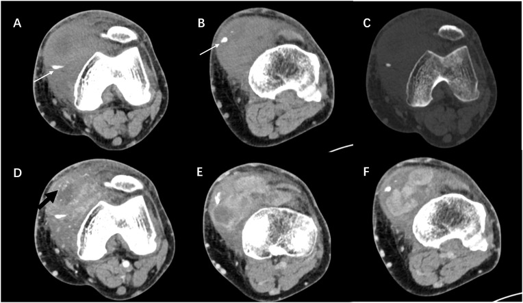
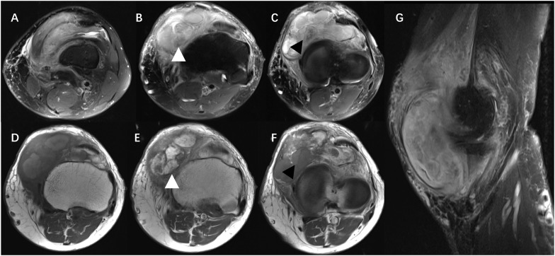
Solitary fibrous tumors (SFTs) are classified as fibroblastic/myofibroblastic tumors that originate from CD34-positive dendritic cells and usually occur in the pleura. In this paper, we describe a case of SFT within the joint cavity of the left knee. A 60-year-old man was admitted to hospital due to swelling in the left knee for the past 8 months without relevant trauma history. X-ray, computed tomography (CT), magnetic resonance imaging (MRI), and positron emission tomography-computed tomography (PET-CT) presented a large, ill-circumscribed, hypervascular, and highly enhanced mass with eccentric calcification and peripheral, intra-lesional vessels. Subsequently, the patient underwent surgical resection. Postoperative pathology confirmed the neoplastic cells to be positive for CD34, Bcl-2, and SATA6, therefore was finally diagnosed as malignant SFT. The patient developed bone metastases within 1 year after surgery. SFT in the joint cavity is rare, and it is difficult to make a preoperative diagnosis.
孤立性纤维性肿瘤(SFTs)被归类为起源于CD34阳性树突状细胞的成纤维细胞/肌成纤维细胞肿瘤,通常发生于胸膜。在本文中,我们描述了一例发生于左膝关节腔内的SFT病例。一名60岁男性因左膝肿胀8个月入院,无相关外伤史。X线、计算机断层扫描(CT)、磁共振成像(MRI)和正电子发射断层扫描-计算机断层扫描(PET-CT)显示为一个大的、边界不清、血管丰富且强化明显的肿块,伴有偏心钙化及外周和瘤内血管。随后,患者接受了手术切除。术后病理证实肿瘤细胞CD34、Bcl-2和SATA6呈阳性,因此最终诊断为恶性SFT。患者术后1年内发生了骨转移。关节腔内的SFT罕见,术前诊断困难。