• 文献检索
  • 文档翻译
  • 深度研究
  • 学术资讯
  • Suppr Zotero 插件Zotero 插件
  • 邀请有礼
  • 套餐&价格
  • 历史记录
应用&插件
Suppr Zotero 插件Zotero 插件浏览器插件Mac 客户端Windows 客户端微信小程序
定价
高级版会员购买积分包购买API积分包
服务
文献检索文档翻译深度研究API 文档MCP 服务
关于我们
关于 Suppr公司介绍联系我们用户协议隐私条款
关注我们

Suppr 超能文献

核心技术专利:CN118964589B侵权必究
粤ICP备2023148730 号-1Suppr @ 2026

文献检索

告别复杂PubMed语法,用中文像聊天一样搜索,搜遍4000万医学文献。AI智能推荐,让科研检索更轻松。

立即免费搜索

文件翻译

保留排版,准确专业,支持PDF/Word/PPT等文件格式,支持 12+语言互译。

免费翻译文档

深度研究

AI帮你快速写综述,25分钟生成高质量综述,智能提取关键信息,辅助科研写作。

立即免费体验

纤连蛋白在浆液性上皮性卵巢癌中的表达有可能阻碍转移扩散和上皮-间质转化:免疫组织化学和电子数据集的比较表达分析

Plakin Expression in Serous Epithelial Ovarian Cancer Has the Potential to Impede Metastatic Spread and Epithelial-Mesenchymal Transition: A Comparative Expression Analysis of Immunohistochemical and In Silico Datasets.

作者信息

Wesley Tamsin, Escalona Ruth M, Kannourakis George, Ahmed Nuzhat

机构信息

Fiona Elsey Cancer Research Institute, Ballarat, VIC 3353, Australia.

Health Innovation and Transformation Centre, Mt Helen Campus, Federation University Australia, Ballarat, VIC 3353, Australia.

出版信息

Cancers (Basel). 2024 Dec 6;16(23):4087. doi: 10.3390/cancers16234087.

DOI:10.3390/cancers16234087
PMID:39682273
原文链接:https://pmc.ncbi.nlm.nih.gov/articles/PMC11639978/
Abstract

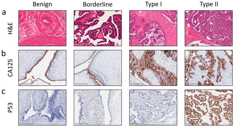
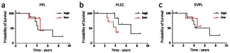
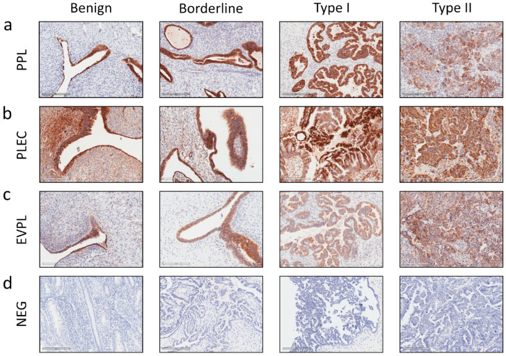
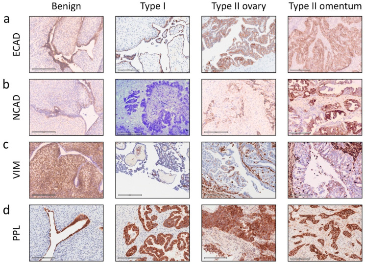
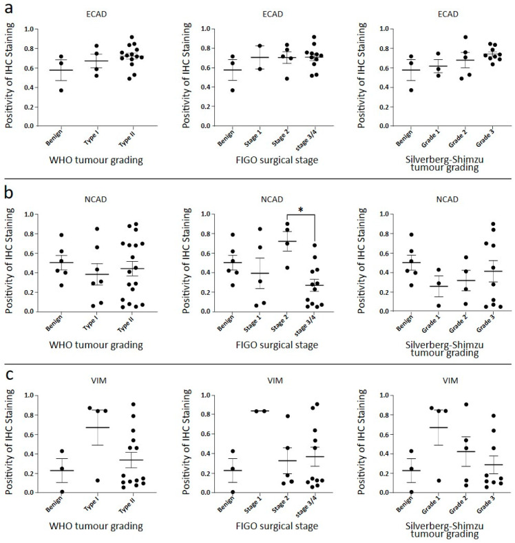
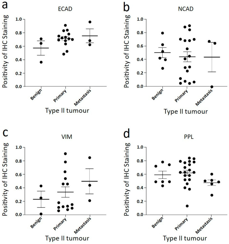
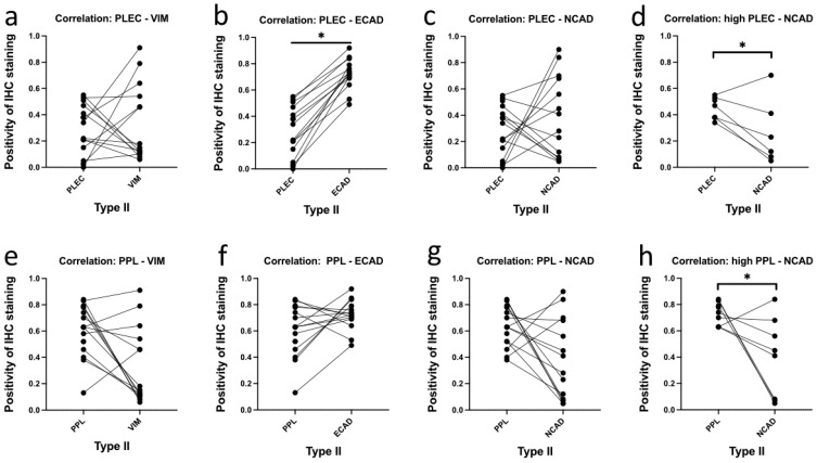
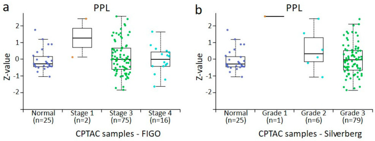
Epithelial ovarian cancer is aggressive and causes high mortality among women worldwide. Members of the plakin family are essential to maintain cytoskeletal integrity and key cellular processes. In this study we characterised the expression of plakins, particularly plectin (PLEC), periplakin (PPL), envoplakin (EVPL), and EMT-related proteins by immunohistochemistry in n = 48 patients' samples to evaluate a potential correlation of plakin expression with EMT as EOC progresses. These tissue plakin and EMT expression analyses were further evaluated by in vitro cell line expression and correlated with the expression of these molecules using publicly available datasets such as Cancer Genome Atlas (TCGA) and Clinical Proteome Tumour Analysis Consortium (CPTAC) datasets. We demonstrate that the expression of PPL and PLEC plakins is decreased in high-grade compared to low-grade EOCs with mixed EMT marker protein expression. This is supported by the correlation of high PPL and PLEC expression with an epithelial rather than mesenchymal phenotype. Our data suggest a partial loss of plakin expression as EOC tumours progress. This may impact the connections of plakins with membrane-bound receptors, which impede the downstream signalling required for the initiation of EMT as the tumours progress.

摘要

上皮性卵巢癌具有侵袭性,在全球女性中导致高死亡率。斑块蛋白家族成员对于维持细胞骨架完整性和关键细胞过程至关重要。在本研究中,我们通过免疫组织化学对48例患者样本中斑块蛋白的表达进行了表征,特别是血影蛋白(PLEC)、周斑蛋白(PPL)、内斑蛋白(EVPL)和与上皮-间质转化(EMT)相关的蛋白,以评估随着上皮性卵巢癌(EOC)进展,斑块蛋白表达与EMT之间的潜在相关性。通过体外细胞系表达进一步评估了这些组织中斑块蛋白和EMT的表达,并使用公开可用的数据集,如癌症基因组图谱(TCGA)和临床蛋白质组肿瘤分析联盟(CPTAC)数据集,将其与这些分子的表达进行关联。我们证明,与低级别EOC相比,高级别EOC中PPL和PLEC斑块蛋白的表达降低,且伴有混合的EMT标记蛋白表达。高PPL和PLEC表达与上皮而非间质表型的相关性支持了这一点。我们的数据表明,随着EOC肿瘤的进展,斑块蛋白表达部分丧失。这可能会影响斑块蛋白与膜结合受体的连接,从而在肿瘤进展时阻碍启动EMT所需的下游信号传导。

https://cdn.ncbi.nlm.nih.gov/pmc/blobs/d293/11639978/9e86831684ec/cancers-16-04087-g019.jpg
https://cdn.ncbi.nlm.nih.gov/pmc/blobs/d293/11639978/096c336d8662/cancers-16-04087-g001.jpg
https://cdn.ncbi.nlm.nih.gov/pmc/blobs/d293/11639978/68c7c0611bd9/cancers-16-04087-g002.jpg
https://cdn.ncbi.nlm.nih.gov/pmc/blobs/d293/11639978/e857462e6a76/cancers-16-04087-g003.jpg
https://cdn.ncbi.nlm.nih.gov/pmc/blobs/d293/11639978/1b141096d1f5/cancers-16-04087-g004.jpg
https://cdn.ncbi.nlm.nih.gov/pmc/blobs/d293/11639978/edd7fc88dc8d/cancers-16-04087-g005.jpg
https://cdn.ncbi.nlm.nih.gov/pmc/blobs/d293/11639978/72f7f4cc14f1/cancers-16-04087-g006.jpg
https://cdn.ncbi.nlm.nih.gov/pmc/blobs/d293/11639978/0f5598c0e948/cancers-16-04087-g007.jpg
https://cdn.ncbi.nlm.nih.gov/pmc/blobs/d293/11639978/dc9b4400c000/cancers-16-04087-g008.jpg
https://cdn.ncbi.nlm.nih.gov/pmc/blobs/d293/11639978/293aaa438f5f/cancers-16-04087-g009.jpg
https://cdn.ncbi.nlm.nih.gov/pmc/blobs/d293/11639978/b4b206b0a4f5/cancers-16-04087-g010.jpg
https://cdn.ncbi.nlm.nih.gov/pmc/blobs/d293/11639978/e6110e7fcde5/cancers-16-04087-g011.jpg
https://cdn.ncbi.nlm.nih.gov/pmc/blobs/d293/11639978/2aafc5c39aee/cancers-16-04087-g012.jpg
https://cdn.ncbi.nlm.nih.gov/pmc/blobs/d293/11639978/3f6e3e25edc4/cancers-16-04087-g013.jpg
https://cdn.ncbi.nlm.nih.gov/pmc/blobs/d293/11639978/1b00c49118c4/cancers-16-04087-g014.jpg
https://cdn.ncbi.nlm.nih.gov/pmc/blobs/d293/11639978/d2ab60a4c9a5/cancers-16-04087-g015.jpg
https://cdn.ncbi.nlm.nih.gov/pmc/blobs/d293/11639978/1c463f5af938/cancers-16-04087-g016.jpg
https://cdn.ncbi.nlm.nih.gov/pmc/blobs/d293/11639978/daa8de0845fd/cancers-16-04087-g017.jpg
https://cdn.ncbi.nlm.nih.gov/pmc/blobs/d293/11639978/bc56d3389346/cancers-16-04087-g018.jpg
https://cdn.ncbi.nlm.nih.gov/pmc/blobs/d293/11639978/9e86831684ec/cancers-16-04087-g019.jpg

相似文献

1
Plakin Expression in Serous Epithelial Ovarian Cancer Has the Potential to Impede Metastatic Spread and Epithelial-Mesenchymal Transition: A Comparative Expression Analysis of Immunohistochemical and In Silico Datasets.纤连蛋白在浆液性上皮性卵巢癌中的表达有可能阻碍转移扩散和上皮-间质转化:免疫组织化学和电子数据集的比较表达分析
Cancers (Basel). 2024 Dec 6;16(23):4087. doi: 10.3390/cancers16234087.
2
The attributes of plakins in cancer and disease: perspectives on ovarian cancer progression, chemoresistance and recurrence. plakins 在癌症和疾病中的特性:对卵巢癌进展、化疗耐药和复发的展望。
Cell Commun Signal. 2021 May 17;19(1):55. doi: 10.1186/s12964-021-00726-x.
3
The members of the plakin family of proteins recognized by paraneoplastic pemphigus antibodies include periplakin.副肿瘤性天疱疮抗体识别的plakin家族蛋白质成员包括周膜蛋白。
J Invest Dermatol. 1998 Aug;111(2):308-13. doi: 10.1046/j.1523-1747.1998.00279.x.
4
Loss of periplakin expression is associated with the tumorigenesis of colorectal carcinoma.外周斑蛋白表达缺失与结直肠癌的肿瘤发生相关。
Biomed Pharmacother. 2017 Mar;87:366-374. doi: 10.1016/j.biopha.2016.12.103. Epub 2017 Jan 6.
5
Unique role for the periplakin tail in intermediate filament association: specific binding to keratin 8 and vimentin.桥粒斑蛋白尾部在中间丝结合中的独特作用:与角蛋白8和波形蛋白的特异性结合。
Exp Dermatol. 2002 Oct;11(5):428-38. doi: 10.1034/j.1600-0625.2002.110506.x.
6
Plakins in development and disease.发育和疾病中的斑联蛋白
Exp Cell Res. 2007 Jun 10;313(10):2189-203. doi: 10.1016/j.yexcr.2007.03.039. Epub 2007 Apr 19.
7
In Vitro and In Silico Analysis of Epithelial-Mesenchymal Transition and Cancer Stemness as Prognostic Markers of Clear Cell Renal Cell Carcinoma.上皮-间质转化和癌症干性作为透明细胞肾细胞癌预后标志物的体外和计算机模拟分析
Cancers (Basel). 2023 May 1;15(9):2586. doi: 10.3390/cancers15092586.
8
Cysteine-Rich Intestinal Protein 1 Served as an Epithelial Ovarian Cancer Marker via Promoting Wnt/-Catenin-Mediated EMT and Tumour Metastasis.富含半胱氨酸的肠内蛋白 1 通过促进 Wnt/-连环蛋白介导的 EMT 和肿瘤转移,作为卵巢癌的上皮标志物。
Dis Markers. 2021 Aug 6;2021:3566749. doi: 10.1155/2021/3566749. eCollection 2021.
9
Functional Analysis of Periplakin and Envoplakin, Cytoskeletal Linkers, and Cornified Envelope Precursor Proteins.外周斑蛋白和内披蛋白的功能分析,细胞骨架连接蛋白及角质包膜前体蛋白
Methods Enzymol. 2016;569:309-29. doi: 10.1016/bs.mie.2015.06.019. Epub 2015 Jul 6.
10
MDM2 promotes epithelial-mesenchymal transition and metastasis of ovarian cancer SKOV3 cells.MDM2促进卵巢癌SKOV3细胞的上皮-间质转化和转移。
Br J Cancer. 2017 Oct 10;117(8):1192-1201. doi: 10.1038/bjc.2017.265. Epub 2017 Aug 17.

引用本文的文献

1
Identification of Proteins Associated with Ovarian Cancer Chemotherapy Resistance Using MALDI-MSI.使用基质辅助激光解吸电离质谱成像技术鉴定与卵巢癌化疗耐药相关的蛋白质
Int J Mol Sci. 2025 Jun 19;26(12):5893. doi: 10.3390/ijms26125893.

本文引用的文献

1
Global cancer statistics 2022: GLOBOCAN estimates of incidence and mortality worldwide for 36 cancers in 185 countries.2022 年全球癌症统计数据:全球 185 个国家和地区 36 种癌症的发病率和死亡率全球估计数。
CA Cancer J Clin. 2024 May-Jun;74(3):229-263. doi: 10.3322/caac.21834. Epub 2024 Apr 4.
2
CXCR4 and CXCR7 signaling promotes tumor progression and obesity-associated epithelial-mesenchymal transition in prostate cancer cells.CXCR4和CXCR7信号传导促进前列腺癌细胞的肿瘤进展和肥胖相关的上皮-间质转化。
Oncogene. 2022 Oct;41(41):4633-4644. doi: 10.1038/s41388-022-02466-9. Epub 2022 Sep 10.
3
Disassembly of α6β4-mediated hemidesmosomal adhesions promotes tumorigenesis in PTEN-negative prostate cancer by targeting plectin to focal adhesions.
α6β4 介导的半桥粒黏附的解体通过将网蛋白靶向黏着斑促进 PTEN 阴性前列腺癌的发生。
Oncogene. 2022 Jul;41(30):3804-3820. doi: 10.1038/s41388-022-02389-5. Epub 2022 Jul 1.
4
MicroRNA-485-5p targets keratin 17 to regulate oral cancer stemness and chemoresistance via the integrin/FAK/Src/ERK/β-catenin pathway.微小 RNA-485-5p 通过整合素/FAK/Src/ERK/β-连环蛋白通路靶向角蛋白 17 调节口腔癌干性和化疗耐药性。
J Biomed Sci. 2022 Jun 15;29(1):42. doi: 10.1186/s12929-022-00824-z.
5
Prognostic implications of PPL expression in ovarian cancer.PPL表达在卵巢癌中的预后意义。
Discov Oncol. 2022 May 25;13(1):35. doi: 10.1007/s12672-022-00496-z.
6
Targeting the PI3K/AKT/mTOR pathway in epithelial ovarian cancer, therapeutic treatment options for platinum-resistant ovarian cancer.靶向上皮性卵巢癌中的PI3K/AKT/mTOR信号通路,铂耐药卵巢癌的治疗选择
Cancer Drug Resist. 2021 Apr 14;4(3):573-595. doi: 10.20517/cdr.2021.05. eCollection 2021.
7
UALCAN: An update to the integrated cancer data analysis platform.UALCAN:一个集成癌症数据分析平台的更新。
Neoplasia. 2022 Mar;25:18-27. doi: 10.1016/j.neo.2022.01.001. Epub 2022 Jan 22.
8
The attributes of plakins in cancer and disease: perspectives on ovarian cancer progression, chemoresistance and recurrence. plakins 在癌症和疾病中的特性:对卵巢癌进展、化疗耐药和复发的展望。
Cell Commun Signal. 2021 May 17;19(1):55. doi: 10.1186/s12964-021-00726-x.
9
What Is New on Ovarian Carcinoma: Integrated Morphologic and Molecular Analysis Following the New 2020 World Health Organization Classification of Female Genital Tumors.卵巢癌的新进展:遵循2020年世界卫生组织女性生殖器官肿瘤新分类的综合形态学和分子分析
Diagnostics (Basel). 2021 Apr 14;11(4):697. doi: 10.3390/diagnostics11040697.
10
Understanding the effect of mechanical forces on ovarian cancer progression.了解机械力对卵巢癌进展的影响。
Gynecol Oncol. 2021 Jul;162(1):154-162. doi: 10.1016/j.ygyno.2021.04.003. Epub 2021 Apr 20.