• 文献检索
  • 文档翻译
  • 深度研究
  • 学术资讯
  • Suppr Zotero 插件Zotero 插件
  • 邀请有礼
  • 套餐&价格
  • 历史记录
应用&插件
Suppr Zotero 插件Zotero 插件浏览器插件Mac 客户端Windows 客户端微信小程序
定价
高级版会员购买积分包购买API积分包
服务
文献检索文档翻译深度研究API 文档MCP 服务
关于我们
关于 Suppr公司介绍联系我们用户协议隐私条款
关注我们

Suppr 超能文献

核心技术专利:CN118964589B侵权必究
粤ICP备2023148730 号-1Suppr @ 2026

文献检索

告别复杂PubMed语法,用中文像聊天一样搜索,搜遍4000万医学文献。AI智能推荐,让科研检索更轻松。

立即免费搜索

文件翻译

保留排版,准确专业,支持PDF/Word/PPT等文件格式,支持 12+语言互译。

免费翻译文档

深度研究

AI帮你快速写综述,25分钟生成高质量综述,智能提取关键信息,辅助科研写作。

立即免费体验

动态高分辨率超声评估颞下颌关节紊乱病中关节盘前移位的诊断准确性:一项前瞻性观察研究

Diagnostic Accuracy of Dynamic High-Resolution Ultrasonography in Assessing Anterior Disc Displacement in Temporomandibular Joint Disorders: A Prospective Observational Study.

作者信息

Wang Kaili, Li Chenyang, Zhou Jinbo, Ren Jiayin, You Meng

机构信息

State Key Laboratory of Oral Diseases, National Center for Stomatology, National Clinical Research Center for Oral Diseases, Department of Oral Medical Imaging, West China Hospital of Stomatology, Sichuan University, Chengdu 610041, China.

出版信息

Healthcare (Basel). 2024 Nov 25;12(23):2355. doi: 10.3390/healthcare12232355.

DOI:10.3390/healthcare12232355
PMID:39684977
原文链接:https://pmc.ncbi.nlm.nih.gov/articles/PMC11641683/
Abstract

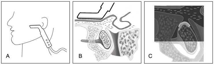
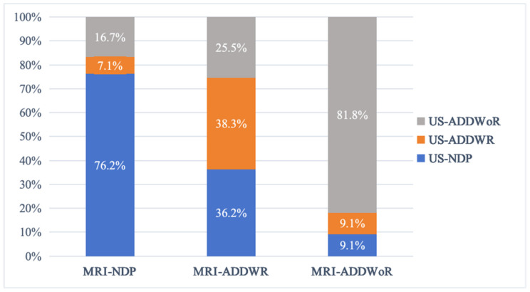
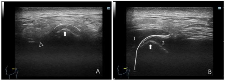
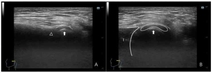
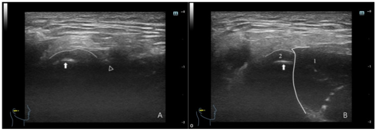
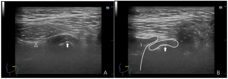
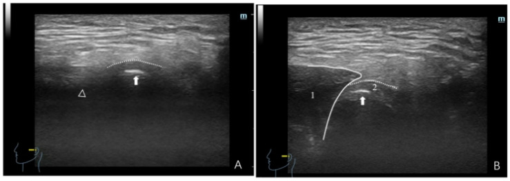
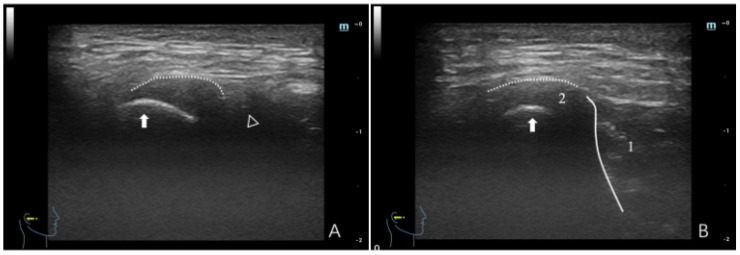
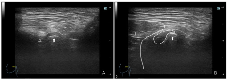
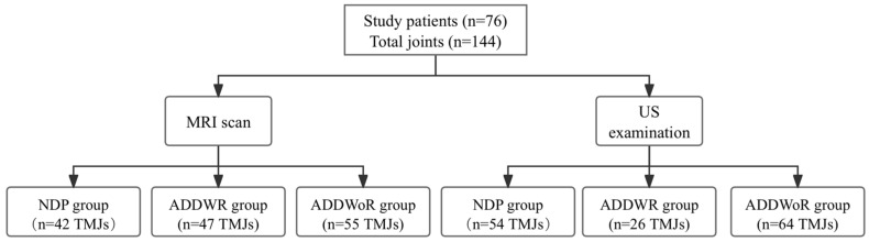
The objective of this study was to assess the diagnostic efficacy of dynamic high-resolution ultrasonography (HRUS) in detecting anterior disc displacement with reduction (ADDWR) and anterior disc displacement without reduction (ADDWoR) in the temporomandibular joint (TMJ). A total of 144 TMJs was categorized into three groups according to the magnetic resonance imaging (MRI) findings, which served as the reference standard: the normal disc position (NDP) group, the ADDWR group, and the ADDWoR group. Static images of the TMJ in full opening and maximum intercuspal positions, along with dynamic sequences during jaw opening, were obtained utilizing a 14 MHz L-shaped linear array transducer. The diagnostic efficacy of dynamic HRUS for identifying ADDWR and ADDWoR was evaluated in terms of accuracy, sensitivity, specificity, positive predictive value (PPV), negative predictive value (NPV), diagnostic odds ratio (DOR), and the Youden index. According to the MRI findings, the NDP, ADDWR, and ADDWoR groups consisted of 42 (29.2%), 47 (32.6%), and 55 (38.2%) TMJs, respectively. HRUS data revealed 54 TMJs (37.5%) in the NDP group, 26 TMJs (18.1%) in the ADDWR group, and 64 TMJs (44.4%) in the ADDWoR group. With MRI as the reference standard, HRUS exhibited a diagnostic accuracy of 71.4%, sensitivity of 51.4%, and specificity of 91.4% for ADDWR. For the ADDWoR, HRUS attained a diagnostic accuracy of 86.5%, sensitivity of 90.0%, and specificity of 82.1%. With MRI serving as the reference standard, dynamic HRUS has high diagnostic value for ADDWoR, with better diagnostic accuracy than ADDWR. Ultrasonography has the potential to be used as a highly effective and non-invasive imaging modality for the early screening of ADD in future clinical practice.

摘要

本研究的目的是评估动态高分辨率超声检查(HRUS)在检测颞下颌关节(TMJ)可复性盘前移位(ADDWR)和不可复性盘前移位(ADDWoR)方面的诊断效能。根据磁共振成像(MRI)结果,将总共144个TMJ分为三组,MRI结果作为参考标准:正常盘位置(NDP)组、ADDWR组和ADDWoR组。使用14 MHz L形线性阵列换能器获取TMJ在最大开口位和最大牙尖交错位的静态图像,以及开口过程中的动态序列。从准确性、敏感性、特异性、阳性预测值(PPV)、阴性预测值(NPV)、诊断比值比(DOR)和尤登指数方面评估动态HRUS对识别ADDWR和ADDWoR的诊断效能。根据MRI结果,NDP组、ADDWR组和ADDWoR组分别由42个(29.2%)、47个(32.6%)和55个(38.2%)TMJ组成。HRUS数据显示,NDP组有54个TMJ(37.5%),ADDWR组有26个TMJ(18.1%),ADDWoR组有64个TMJ(44.4%)。以MRI作为参考标准,HRUS对ADDWR的诊断准确性为71.4%,敏感性为51.4%,特异性为91.4%。对于ADDWoR,HRUS的诊断准确性为86.5%,敏感性为90.0%,特异性为82.1%。以MRI作为参考标准,动态HRUS对ADDWoR具有较高的诊断价值,诊断准确性优于ADDWR。超声检查有可能在未来临床实践中作为一种高效且无创的成像方式用于ADD的早期筛查。

https://cdn.ncbi.nlm.nih.gov/pmc/blobs/5cf3/11641683/5ec2a4e39000/healthcare-12-02355-g010.jpg
https://cdn.ncbi.nlm.nih.gov/pmc/blobs/5cf3/11641683/0bf3b2e84ee6/healthcare-12-02355-g001.jpg
https://cdn.ncbi.nlm.nih.gov/pmc/blobs/5cf3/11641683/f193f72cad0d/healthcare-12-02355-g002.jpg
https://cdn.ncbi.nlm.nih.gov/pmc/blobs/5cf3/11641683/a91e50219249/healthcare-12-02355-g003.jpg
https://cdn.ncbi.nlm.nih.gov/pmc/blobs/5cf3/11641683/9a940a6b201d/healthcare-12-02355-g004.jpg
https://cdn.ncbi.nlm.nih.gov/pmc/blobs/5cf3/11641683/b41d3b013651/healthcare-12-02355-g005.jpg
https://cdn.ncbi.nlm.nih.gov/pmc/blobs/5cf3/11641683/af7d9d1dfdf7/healthcare-12-02355-g006.jpg
https://cdn.ncbi.nlm.nih.gov/pmc/blobs/5cf3/11641683/bc9c0952b103/healthcare-12-02355-g007.jpg
https://cdn.ncbi.nlm.nih.gov/pmc/blobs/5cf3/11641683/61bb4067b741/healthcare-12-02355-g008.jpg
https://cdn.ncbi.nlm.nih.gov/pmc/blobs/5cf3/11641683/a0b6896f2b4e/healthcare-12-02355-g009.jpg
https://cdn.ncbi.nlm.nih.gov/pmc/blobs/5cf3/11641683/5ec2a4e39000/healthcare-12-02355-g010.jpg

相似文献

1
Diagnostic Accuracy of Dynamic High-Resolution Ultrasonography in Assessing Anterior Disc Displacement in Temporomandibular Joint Disorders: A Prospective Observational Study.动态高分辨率超声评估颞下颌关节紊乱病中关节盘前移位的诊断准确性:一项前瞻性观察研究
Healthcare (Basel). 2024 Nov 25;12(23):2355. doi: 10.3390/healthcare12232355.
2
[A three-dimensional imaging study of the effect of anterior disc displacement on the bony structural relationship of the temporomandibular joint].[关节盘前移位对颞下颌关节骨结构关系影响的三维成像研究]
Zhonghua Kou Qiang Yi Xue Za Zhi. 2024 Feb 9;59(2):157-164. doi: 10.3760/cma.j.cn112144-20230718-00015.
3
[The diagnostic value of high-resolution ultrasonography for detecting anterior disc displacement without reduction of temporomandibular joint].[高分辨率超声对颞下颌关节不可复性盘前移位的诊断价值]
Hua Xi Kou Qiang Yi Xue Za Zhi. 2012 Dec;30(6):632-4, 640.
4
Role of Dynamic 3 Tesla MRI in the Evaluation of Temporomandibular Joint Dysfunction.动态3特斯拉磁共振成像在颞下颌关节紊乱评估中的作用
Cureus. 2023 Mar 25;15(3):e36681. doi: 10.7759/cureus.36681. eCollection 2023 Mar.
5
Evaluation of clinical signs and magnetic resonance imaging findings in patients with temporomandibular disorders.颞下颌关节紊乱病患者临床体征及磁共振成像结果评估
J Craniomaxillofac Surg. 2023 Jul-Aug;51(7-8):441-447. doi: 10.1016/j.jcms.2023.08.013. Epub 2023 Aug 16.
6
Comparison of altered signal intensity, position, and morphology of the TMJ disc in MR images corrected for variations in surface coil sensitivity.针对表面线圈灵敏度变化进行校正后的磁共振图像中颞下颌关节盘信号强度、位置及形态改变的比较。
Oral Surg Oral Med Oral Pathol Oral Radiol Endod. 2006 Apr;101(4):515-22. doi: 10.1016/j.tripleo.2005.04.004. Epub 2005 Sep 19.
7
The diagnostic value of high-resolution ultrasonography for the detection of anterior disc displacement of the temporomandibular joint: a meta-analysis employing the HSROC statistical model.高分辨率超声检查对颞下颌关节盘前移位检测的诊断价值:采用HSROC统计模型的Meta分析
Int J Oral Maxillofac Surg. 2015 Jul;44(7):852-8. doi: 10.1016/j.ijom.2015.01.012. Epub 2015 Feb 20.
8
Investigation of Morphologic Changes in Temporomandibular Joint With Anterior Disk Displacement Based on 3-Dimensional Reconstructive Imaging.基于三维重建成像的颞下颌关节前盘移位的形态学变化研究。
J Craniofac Surg. 2023 Jun 1;34(4):1271-1277. doi: 10.1097/SCS.0000000000009139. Epub 2022 Nov 28.
9
Association Between Articular Eminence Structure and Joint Spaces in Temporomandibular Joints with Anterior Disc Displacement.关节突形态与伴前移位盘的 TMJ 关节间隙的关系
Niger J Clin Pract. 2024 Mar 1;27(3):408-414. doi: 10.4103/njcp.njcp_710_23. Epub 2024 Mar 26.
10
Relationship between temporomandibular joint space and articular disc displacement.颞下颌关节间隙与关节盘移位之间的关系。
BMC Oral Health. 2025 Apr 21;25(1):611. doi: 10.1186/s12903-025-05991-7.

引用本文的文献

1
Recent advances and educational strategies in diagnostic imaging for temporomandibular disorders: a narrative literature review.颞下颌关节紊乱病诊断成像的最新进展及教育策略:一篇叙述性文献综述
Front Neurol. 2025 May 26;16:1597312. doi: 10.3389/fneur.2025.1597312. eCollection 2025.

本文引用的文献

1
Current status of dynamic musculoskeletal ultrasound for application to treatment of orthopedic diseases.动态肌肉骨骼超声在骨科疾病治疗中的应用现状
Am J Transl Res. 2024 Jun 15;16(6):2180-2189. doi: 10.62347/WHER3512. eCollection 2024.
2
Diagnostic efficacy of quantitative ultrasonography for anterior disc displacement of the temporomandibular joint.定量超声检查对颞下颌关节盘前移位的诊断效能
Quant Imaging Med Surg. 2023 Oct 1;13(10):6446-6455. doi: 10.21037/qims-23-401. Epub 2023 Aug 3.
3
Diagnostic accuracy of ultrasonography compared with magnetic resonance imaging in diagnosing disc displacement of the temporomandibular joint: A systematic review and meta-analysis.
超声检查与磁共振成像在诊断颞下颌关节盘移位中的诊断准确性:一项系统评价和荟萃分析。
J Prosthet Dent. 2025 Feb;133(2):446-454. doi: 10.1016/j.prosdent.2023.03.012. Epub 2023 Apr 17.
4
Advantages of ultrasound guidance for TMJ arthrocentesis and intra-articular injection: A narrative review.超声引导下颞下颌关节穿刺术和关节内注射的优势:一项叙述性综述。
Dent Med Probl. 2022 Oct-Dec;59(4):647-656. doi: 10.17219/dmp/146820.
5
Diagnosis of temporomandibular disorders using artificial intelligence technologies: A systematic review and meta-analysis.使用人工智能技术诊断颞下颌关节紊乱:系统评价和荟萃分析。
PLoS One. 2022 Aug 18;17(8):e0272715. doi: 10.1371/journal.pone.0272715. eCollection 2022.
6
Temporomandibular joint arthrocentesis guided by ultrasonography: An anatomical study.超声引导下颞下颌关节关节腔穿刺术:一项解剖学研究。
J Stomatol Oral Maxillofac Surg. 2021 Sep;122(4):e27-e31. doi: 10.1016/j.jormas.2021.03.002. Epub 2021 Mar 8.
7
Ultrasound-guided vs conventional arthrocentesis for management of temporomandibular joint disorders: A systematic review and meta-analysis.超声引导下与常规关节腔穿刺术治疗颞下颌关节紊乱病的比较:系统评价和荟萃分析。
Cranio. 2023 May;41(3):264-273. doi: 10.1080/08869634.2020.1829870. Epub 2020 Oct 12.
8
Imaging of the temporomandibular joint.颞下颌关节影像学。
Clin Radiol. 2021 Jan;76(1):76.e21-76.e35. doi: 10.1016/j.crad.2020.06.020. Epub 2020 Jul 21.
9
Comparison of clinical and magnetic resonance imagining data of patients with temporomandibular disorders.颞下颌关节紊乱病患者的临床与磁共振成像数据比较。
Niger J Clin Pract. 2020 Mar;23(3):376-380. doi: 10.4103/njcp.njcp_492_19.
10
Treatment of Temporomandibular Disorders: Symptoms Versus Cause.
J Oral Maxillofac Surg. 2020 May;78(5):675. doi: 10.1016/j.joms.2019.12.032. Epub 2020 Jan 12.