Yamada Takayuki, Yanagaki Satoru, Satani Nozomi, Kagaya Yuriko, Sato Tomoni, Matsuura Tomonori, Sato Teruyuki, Noguchi Naoya, Ohta Nobuo
Department of Radiology, Tohoku Medical and Pharmaceutical University, Sendai, Japan.
Department of Otolaryngology, Tohoku Medical and Pharmaceutical University, Sendai, Japan.
Radiol Case Rep. 2024 Nov 30;20(2):1145-1149. doi: 10.1016/j.radcr.2024.11.014. eCollection 2025 Feb.
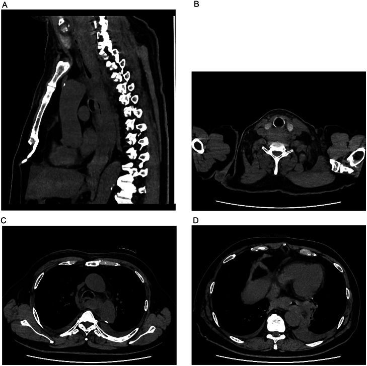
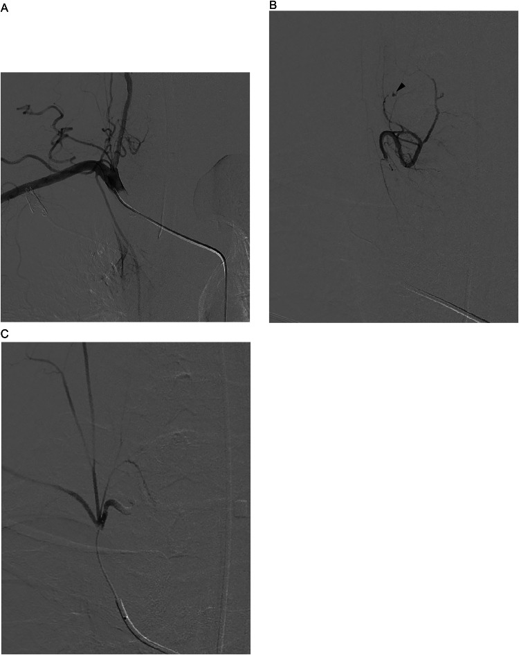
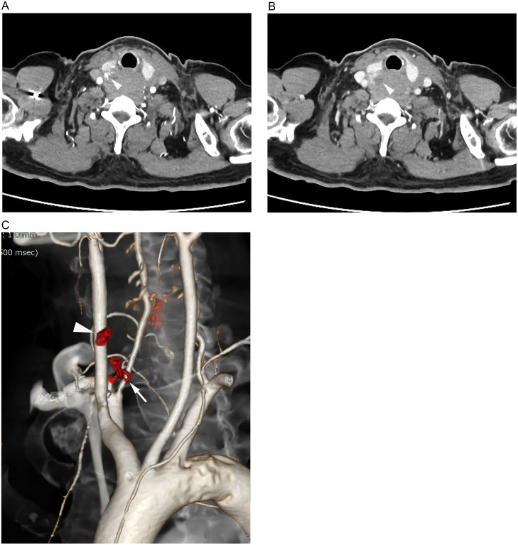
A 62-year-old man was referred to our hospital presenting with a sore throat, dyspnea, and cervical swelling. Initial precontrast CT scans revealed a cervical and mediastinal hematoma, along with a hemothorax. Further dynamic contrast-enhanced CT scans indicated contrast media extravasation dorsal to the right thyroid gland lobe, suggesting a rupture of the right inferior thyroid artery or a parathyroid adenoma. Following endotracheal intubation, angiography confirmed extravasation from the right inferior thyroid artery. Transarterial embolization (TAE) was successfully performed using a gelatin sponge. The cervical and mediastinal hematoma were surgically excised, and the right inferior parathyroid gland was simultaneously resected. Pathological examination revealed no neoplastic components.
一名62岁男性因咽痛、呼吸困难和颈部肿胀被转诊至我院。初始平扫CT扫描显示颈部和纵隔血肿,以及血胸。进一步的动态增强CT扫描显示右甲状腺叶背侧有造影剂外渗,提示右甲状腺下动脉破裂或甲状旁腺腺瘤。气管插管后,血管造影证实造影剂从右甲状腺下动脉外渗。使用明胶海绵成功进行了经动脉栓塞术(TAE)。手术切除了颈部和纵隔血肿,并同时切除了右下甲状旁腺。病理检查未发现肿瘤成分。