Ryu Min Hyung, Yun Jeong H, Kim Kangjin, Gentili Michele, Ghosh Auyon, Sciurba Frank, Barwick Lucas, Limper Andrew, Criner Gerard, Brown Kevin K, Wise Robert, Martinez Fernando J, Flaherty Kevin R, Cho Michael H, Castaldi Peter J, DeMeo Dawn L, Silverman Edwin K, Hersh Craig P, Morrow Jarrett D
Channing Division of Network Medicine, Department of Medicine, Brigham and Women's Hospital, Boston, USA, 181 Longwood Ave, 02115, MA.
Harvard Medical School, Boston, MA, USA.
BMC Genomics. 2024 Dec 18;25(1):1192. doi: 10.1186/s12864-024-11031-5.
Chronic obstructive pulmonary disease (COPD) and idiopathic pulmonary fibrosis (IPF) are debilitating diseases associated with divergent histopathological changes in the lungs. At present, due to cost and technical limitations, profiling cell types is not practical in large epidemiology cohorts (n > 1000). Here, we used computational deconvolution to identify cell types in COPD and IPF lungs whose abundances and cell type-specific gene expression are associated with disease diagnosis and severity.
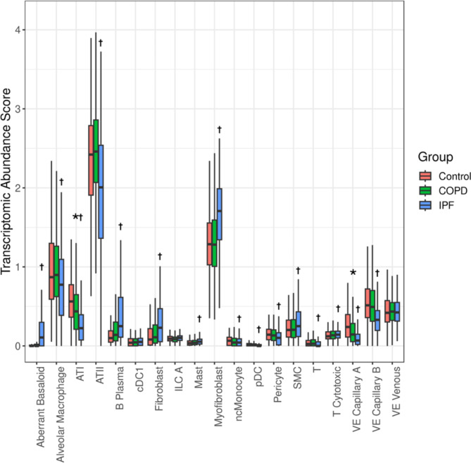
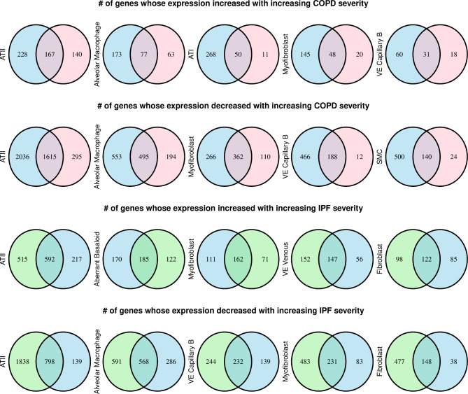
We analyzed lung tissue RNA-seq data from 1026 subjects (COPD, n = 465; IPF, n = 213; control, n = 348) from the Lung Tissue Research Consortium. We performed RNA-seq deconvolution, querying thirty-eight discrete cell-type varieties in the lungs. We tested whether deconvoluted cell-type abundance and cell type-specific gene expression were associated with disease severity. The abundance score of twenty cell types significantly differed between IPF and control lungs. In IPF subjects, eleven and nine cell types were significantly associated with forced vital capacity (FVC) and diffusing capacity for carbon monoxide (DCO), respectively. Aberrant basaloid cells, a rare cells found in fibrotic lungs, were associated with worse FVC and DCO in IPF subjects, indicating that this aberrant epithelial population increased with disease severity. Alveolar type 1 and vascular endothelial (VE) capillary A were decreased in COPD lungs compared to controls. An increase in macrophages and classical monocytes was associated with lower DCO in IPF and COPD subjects. In both diseases, lower non-classical monocytes and VE capillary A cells were associated with increased disease severity. Alveolar type 2 cells and alveolar macrophages had the highest number of genes with cell type-specific differential expression by disease severity in COPD and IPF. In IPF, genes implicated in the pathogenesis of IPF, such as matrix metallopeptidase 7, growth differentiation factor 15, and eph receptor B2, were associated with disease severity in a cell type-specific manner.
Utilization of RNA-seq deconvolution enabled us to pinpoint cell types present in the lungs that are associated with the severity of COPD and IPF. This knowledge offers valuable insight into the alterations within tissues in more advanced illness, ultimately providing a better understanding of the underlying pathological processes that drive disease progression.
慢性阻塞性肺疾病(COPD)和特发性肺纤维化(IPF)是与肺部不同组织病理学变化相关的使人衰弱的疾病。目前,由于成本和技术限制,在大型流行病学队列(n>1000)中分析细胞类型并不实际。在此,我们使用计算反卷积来识别COPD和IPF肺中的细胞类型,其丰度和细胞类型特异性基因表达与疾病诊断和严重程度相关。
我们分析了来自肺组织研究联盟的1026名受试者(COPD,n = 465;IPF,n = 213;对照,n = 348)的肺组织RNA测序数据。我们进行了RNA测序反卷积,查询了肺中38种离散的细胞类型。我们测试了反卷积后的细胞类型丰度和细胞类型特异性基因表达是否与疾病严重程度相关。IPF和对照肺之间20种细胞类型的丰度得分存在显著差异。在IPF受试者中,分别有11种和9种细胞类型与用力肺活量(FVC)和一氧化碳弥散量(DCO)显著相关。异常基底样细胞是在纤维化肺中发现的罕见细胞,在IPF受试者中与较差的FVC和DCO相关,表明这种异常上皮细胞群体随疾病严重程度增加。与对照组相比,COPD肺中的1型肺泡细胞和血管内皮(VE)毛细血管A减少。巨噬细胞和经典单核细胞的增加与IPF和COPD受试者较低的DCO相关。在这两种疾病中,较低的非经典单核细胞和VE毛细血管A细胞与疾病严重程度增加相关。在COPD和IPF中,2型肺泡细胞和肺泡巨噬细胞中因疾病严重程度而具有细胞类型特异性差异表达的基因数量最多。在IPF中,与IPF发病机制相关的基因,如基质金属肽酶7、生长分化因子15和Eph受体B2,以细胞类型特异性方式与疾病严重程度相关。
利用RNA测序反卷积使我们能够确定肺中存在的与COPD和IPF严重程度相关的细胞类型。这一知识为更晚期疾病中组织内的变化提供了有价值的见解,最终有助于更好地理解驱动疾病进展的潜在病理过程。